9BU1 | pdb_00009bu1

Estrogen Receptor Alpha Ligand Binding Domain Y537S Mutant in Complex with (6'-hydroxy-1'-(4-((1-propylazetidin-3-yl)methoxy)phenyl)-1',4'-dihydro-2'H-spiro[cyclopropane-1,3'-isoquinolin]-2'-yl)(phenyl)methanone


Experimental Data Snapshot

  • Method: X-RAY DIFFRACTION
  • Resolution: 1.75 Å
  • R-Value Free: 
    0.226 (Depositor), 0.230 (DCC) 
  • R-Value Work: 
    0.194 (Depositor), 0.200 (DCC) 
  • R-Value Observed: 
    0.196 (Depositor) 

Starting Model: experimental
View more details

wwPDB Validation   3D Report Full Report


Ligand Structure Quality Assessment 

Created with Raphaël 2.3.0Worse 01 BetterLigand structure goodness of fit to experimental dataBest fitted A1ASNClick on this verticalbar to view details

This is version 1.1 of the entry. See complete history


Literature

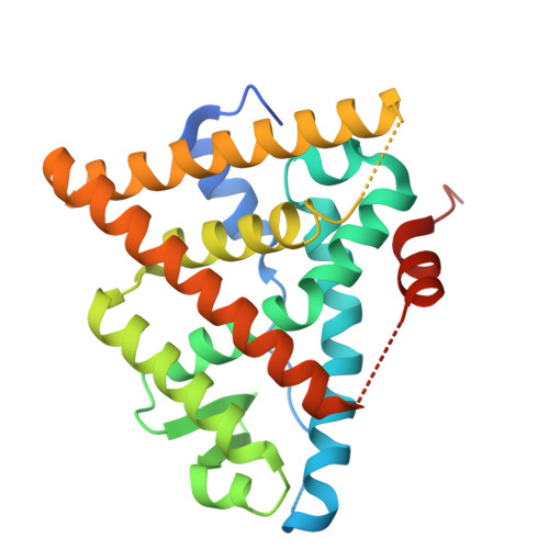
Targeting unique ligand binding domain structural features downregulates DKK1 in Y537S ESR1 mutant breast cancer cells.

Young, K.S.Hancock, G.R.Fink, E.C.Zigrossi, A.Flowers, B.Cooper, D.A.Nguyen, V.T.Martinez, M.C.Mon, K.S.Bosland, M.Zak, D.R.Runde, A.P.Sharifi, M.N.Kastrati, I.Minh, D.D.L.Kregel, S.Fanning, S.W.

(2025) Breast Cancer Res 27: 10-10

  • DOI: https://doi.org/10.1186/s13058-024-01945-z
  • Primary Citation of Related Structures:  
    9BPX, 9BQE, 9BU1

  • PubMed Abstract: 

    Resistance to endocrine therapies remains a major clinical hurdle in breast cancer. Mutations to estrogen receptor alpha (ERα) arise after continued therapeutic pressure. Next generation selective estrogen receptor modulators and degraders/downregulators (SERMs and SERDs) show clinical efficacy, but responses are often non-durable. A tyrosine to serine point mutation at position 537 in the ERα ligand binding domain (LBD) is among the most common and most pathogenic alteration in this setting. It enables endocrine therapy resistance by superceding intrinsic structural-energetic gatekeepers of ER hormone-dependence, it enhances metastatic burden by enabling neomorphic ER-dependent transcriptional programs, and it resists SERM and SERD inhibiton by reducing their binding affinities and abilities to antagonize transcriptional coregulator binding. However, a subset of SERMs and SERDs can achieve efficacy by adopting poses that force the mutation to engage in a new interaction that favors the therapeutic receptor antagonist conformation. We previously described a chemically unconventional SERM, T6I-29, that demonstrates significant anti-proliferative activities in Y537S ERα breast cancer cells. Here, we use a comprehensive suite of structural-biochemical, in vitro, and in vivo approaches to better T6I-29's activities in breast cancer cells harboring Y537S ERα. RNA sequencing in cells treated with T6I-29 reveals a neomorphic downregulation of DKK1, a secreted glycoprotein known to play oncogenic roles in other cancers. Importantly, we find that DKK1 is significantly enriched in ER + breast cancer plasma compared to healthy controls. This study shows how new SERMs and SERDs can identify new therapeutic pathways in endocrine-resistant ER + breast cancers.


  • Organizational Affiliation

    Department of Cancer Biology, Loyola University Chicago Stritch School of Medicine, Maywood, IL, 50153, USA.


Macromolecules
Find similar proteins by:  (by identity cutoff)  |  3D Structure
Entity ID: 1
MoleculeChains Sequence LengthOrganismDetailsImage
Estrogen receptor
A, B
263Homo sapiensMutation(s): 4 
Gene Names: ESR1ESRNR3A1
UniProt & NIH Common Fund Data Resources
Find proteins for P03372 (Homo sapiens)
Explore P03372 
Go to UniProtKB:  P03372
PHAROS:  P03372
GTEx:  ENSG00000091831 
Entity Groups  
Sequence Clusters30% Identity50% Identity70% Identity90% Identity95% Identity100% Identity
UniProt GroupP03372
Sequence Annotations
Expand
  • Reference Sequence
Small Molecules
Ligands 1 Unique
IDChains Name / Formula / InChI Key2D Diagram3D Interactions
A1ASN (Subject of Investigation/LOI)
Query on A1ASN

Download Ideal Coordinates CCD File 
C [auth A],
D [auth B]
[(1'R)-6'-hydroxy-1'-{4-[(1-propylazetidin-3-yl)methoxy]phenyl}-1',4'-dihydro-2'H-spiro[cyclopropane-1,3'-isoquinolin]-2'-yl](phenyl)methanone
C31 H34 N2 O3
LOIMCQIANZASAV-GDLZYMKVSA-N
Experimental Data & Validation

Experimental Data

  • Method: X-RAY DIFFRACTION
  • Resolution: 1.75 Å
  • R-Value Free:  0.226 (Depositor), 0.230 (DCC) 
  • R-Value Work:  0.194 (Depositor), 0.200 (DCC) 
  • R-Value Observed: 0.196 (Depositor) 
Space Group: C 1 2 1
Unit Cell:
Length ( Å )Angle ( ˚ )
a = 102.749α = 90
b = 57.984β = 103.95
c = 87.621γ = 90
Software Package:
Software NamePurpose
PHENIXrefinement
HKL-3000data reduction
HKL-3000data scaling
PHENIXphasing

Structure Validation

View Full Validation Report


Ligand Structure Quality Assessment 

Created with Raphaël 2.3.0Worse 01 BetterLigand structure goodness of fit to experimental dataBest fitted A1ASNClick on this verticalbar to view details

Entry History & Funding Information

Deposition Data


Funding OrganizationLocationGrant Number
National Institutes of Health/National Cancer Institute (NIH/NCI)United States5R37CA279341

Revision History  (Full details and data files)

  • Version 1.0: 2024-06-19
    Type: Initial release
  • Version 1.1: 2025-01-29
    Changes: Database references, Structure summary