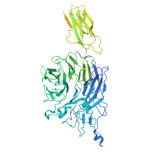
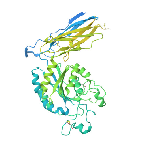
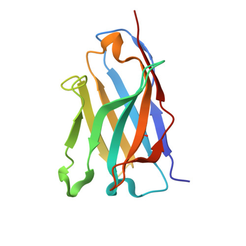
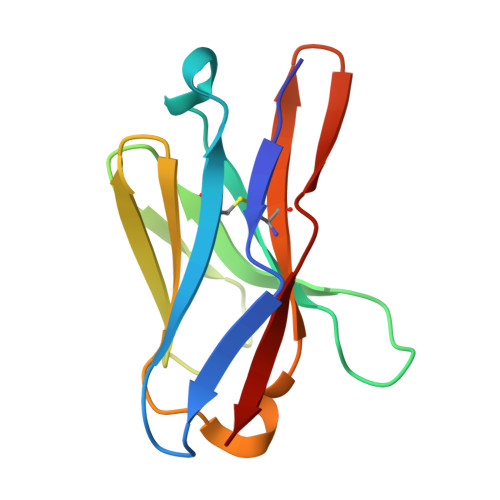
9B9K | pdb_00009b9k

Integrin alpha-5 beta-1 in complex with MINT1526A Fab


Experimental Data Snapshot

  • Method: ELECTRON MICROSCOPY
  • Resolution: 2.70 Å
  • Aggregation State: PARTICLE 
  • Reconstruction Method: SINGLE PARTICLE 

Starting Model: experimental
View more details

wwPDB Validation   3D Report Full Report


This is version 1.0 of the entry. See complete history


Literature

Structural and functional characterization of integrin alpha 5-targeting antibodies for anti-angiogenic therapy.

Nguyen, A.Heim, J.B.Cordara, G.Chan, M.C.Johannesen, H.Charlesworth, C.Li, M.Azumaya, C.M.Madden, B.Krengel, U.Meves, A.Campbell, M.G.

(2025) bioRxiv 

  • DOI: https://doi.org/10.1101/2025.01.08.631572
  • Primary Citation of Related Structures:  
    8R38, 9B9J, 9B9K

  • PubMed Abstract: 

    Integrins are a large family of heterodimeric receptors important for cell adhesion and signaling. Integrin α5β1, also known as the fibronectin receptor, is a key mediator of angiogenesis and its dysregulation is associated with tumor proliferation, progression, and metastasis. Despite numerous efforts, α5β1-targeting therapeutics have been unsuccessful in large part due to efficacy and off-target effects. To mediate activation and signaling, integrins undergo drastic conformational changes. However, how therapeutics influence or are affected by integrin conformation remains incompletely characterized. Using cell biology, biophysics, and electron microscopy, we shed light on these relationships by characterizing two potentially therapeutic anti-α5β1 antibodies, BIIG2 and MINT1526A. We show that both antibodies bind α5β1 with nanomolar affinity and reduce angiogenesis in vitro . We demonstrate BIIG2 reduces tumor growth in two human xenograft mouse models and exhibits a strong specificity for connective tissue-resident fibroblasts and melanoma cells. Using electron microscopy, we map out the molecular interfaces mediating the integrin-antibody interactions and reveal that although both antibodies have overlapping epitopes and block fibronectin binding via steric hindrance, the effect on the conformational equilibrium is drastically different. While MINT1526A constricts α5β1's range of flexibility, BIIG2 binds without restricting the available conformational states. These mechanistic insights, coupled with the functional analysis, guide which aspects should be prioritized to avoid off-target effects or partial agonism in the design of future integrin-targeted therapeutics.


  • Organizational Affiliation

    Basic Sciences Division, Fred Hutchinson Cancer Center, Seattle, Washington 98109, USA.


Macromolecules
Find similar proteins by:  (by identity cutoff)  |  3D Structure
Entity ID: 1
MoleculeChains Sequence LengthOrganismDetailsImage
Integrin alpha-5 light chain995Homo sapiensMutation(s): 0 
Gene Names: ITGA5FNRA
UniProt & NIH Common Fund Data Resources
Find proteins for P08648 (Homo sapiens)
Explore P08648 
Go to UniProtKB:  P08648
PHAROS:  P08648
GTEx:  ENSG00000161638 
Entity Groups  
Sequence Clusters30% Identity50% Identity70% Identity90% Identity95% Identity100% Identity
UniProt GroupP08648
Glycosylation
Glycosylation Sites: 6Go to GlyGen: P08648-1
Sequence Annotations
Expand
  • Reference Sequence
Find similar proteins by:  (by identity cutoff)  |  3D Structure
Entity ID: 2
MoleculeChains Sequence LengthOrganismDetailsImage
Integrin beta-1728Homo sapiensMutation(s): 0 
Gene Names: ITGB1FNRBMDF2MSK12
UniProt & NIH Common Fund Data Resources
Find proteins for P05556 (Homo sapiens)
Explore P05556 
Go to UniProtKB:  P05556
PHAROS:  P05556
GTEx:  ENSG00000150093 
Entity Groups  
Sequence Clusters30% Identity50% Identity70% Identity90% Identity95% Identity100% Identity
UniProt GroupP05556
Glycosylation
Glycosylation Sites: 5Go to GlyGen: P05556-1
Sequence Annotations
Expand
  • Reference Sequence
Find similar proteins by:  (by identity cutoff)  |  3D Structure
Entity ID: 3
MoleculeChains Sequence LengthOrganismDetailsImage
MINT1526A Fab Heavy ChainC [auth H]120unidentifiedMutation(s): 0 
Entity Groups  
Sequence Clusters30% Identity50% Identity70% Identity90% Identity95% Identity100% Identity
Sequence Annotations
Expand
  • Reference Sequence
Find similar proteins by:  (by identity cutoff)  |  3D Structure
Entity ID: 4
MoleculeChains Sequence LengthOrganismDetailsImage
MINT1526A Fab Light ChainD [auth L]112unidentifiedMutation(s): 0 
Entity Groups  
Sequence Clusters30% Identity50% Identity70% Identity90% Identity95% Identity100% Identity
Sequence Annotations
Expand
  • Reference Sequence
Oligosaccharides

Help

Entity ID: 5
MoleculeChains Length2D Diagram Glycosylation3D Interactions
beta-D-mannopyranose-(1-4)-2-acetamido-2-deoxy-beta-D-glucopyranose-(1-4)-2-acetamido-2-deoxy-beta-D-glucopyranoseE [auth C],
G [auth E],
J [auth I],
N
3N-Glycosylation
Glycosylation Resources
GlyTouCan:  G15407YE
GlyCosmos:  G15407YE
GlyGen:  G15407YE
Entity ID: 6
MoleculeChains Length2D Diagram Glycosylation3D Interactions
alpha-D-mannopyranose-(1-3)-[alpha-D-mannopyranose-(1-6)]alpha-D-mannopyranose-(1-6)-[alpha-D-mannopyranose-(1-3)]beta-D-mannopyranose-(1-4)-2-acetamido-2-deoxy-beta-D-glucopyranose-(1-4)-2-acetamido-2-deoxy-beta-D-glucopyranoseF [auth D]7N-Glycosylation
Glycosylation Resources
GlyTouCan:  G55220VL
GlyCosmos:  G55220VL
GlyGen:  G55220VL
Entity ID: 7
MoleculeChains Length2D Diagram Glycosylation3D Interactions
alpha-D-mannopyranose-(1-3)-beta-D-mannopyranose-(1-4)-2-acetamido-2-deoxy-beta-D-glucopyranose-(1-4)-2-acetamido-2-deoxy-beta-D-glucopyranoseH [auth F]4N-Glycosylation
Glycosylation Resources
GlyTouCan:  G81315DD
GlyCosmos:  G81315DD
GlyGen:  G81315DD
Entity ID: 8
MoleculeChains Length2D Diagram Glycosylation3D Interactions
2-acetamido-2-deoxy-beta-D-glucopyranose-(1-4)-2-acetamido-2-deoxy-beta-D-glucopyranoseI [auth G],
K [auth J],
L [auth K],
M
2N-Glycosylation
Glycosylation Resources
GlyTouCan:  G42666HT
GlyCosmos:  G42666HT
GlyGen:  G42666HT
Small Molecules
Ligands 2 Unique
IDChains Name / Formula / InChI Key2D Diagram3D Interactions
NAG
Query on NAG

Download Ideal Coordinates CCD File 
V [auth B]2-acetamido-2-deoxy-beta-D-glucopyranose
C8 H15 N O6
OVRNDRQMDRJTHS-FMDGEEDCSA-N
CA
Query on CA

Download Ideal Coordinates CCD File 
O [auth A]
P [auth A]
Q [auth A]
R [auth A]
S [auth B]
CALCIUM ION
Ca
BHPQYMZQTOCNFJ-UHFFFAOYSA-N
Experimental Data & Validation

Experimental Data

  • Method: ELECTRON MICROSCOPY
  • Resolution: 2.70 Å
  • Aggregation State: PARTICLE 
  • Reconstruction Method: SINGLE PARTICLE 
EM Software:
TaskSoftware PackageVersion
RECONSTRUCTIONcryoSPARC
MODEL REFINEMENTPHENIX
MODEL REFINEMENTISOLDE
MODEL REFINEMENTCoot

Structure Validation

View Full Validation Report


Entry History & Funding Information

Deposition Data


Funding OrganizationLocationGrant Number
National Institutes of Health/National Institute of General Medical Sciences (NIH/NIGMS)United StatesR35 GM147414-01
The Pew Charitable TrustsUnited StatesNA
Other privateP30 CA015704-40

Revision History  (Full details and data files)

  • Version 1.0: 2025-02-05
    Type: Initial release