8R38 | pdb_00008r38

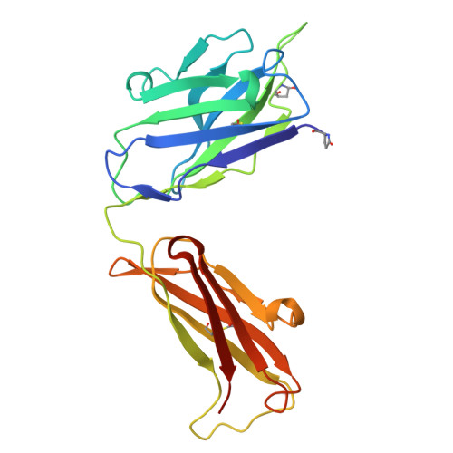
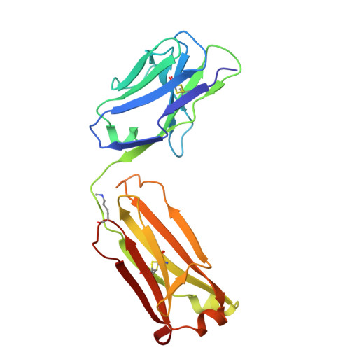
BIIG2 anti-integrin Fab


Experimental Data Snapshot

  • Method: X-RAY DIFFRACTION
  • Resolution: 1.38 Å
  • R-Value Free: 
    0.217 (Depositor), 0.222 (DCC) 
  • R-Value Work: 
    0.195 (Depositor), 0.200 (DCC) 
  • R-Value Observed: 
    0.197 (Depositor) 

Starting Model: experimental
View more details

wwPDB Validation 3D Report Full Report

Validation slider image for 8R38

This is version 1.2 of the entry. See complete history

Literature

Shared ligand-blocking mechanism but distinct conformational modulation by alpha 5-targeting antibodies BIIG2 and MINT1526A.

Nguyen, A.Heim, J.B.Cordara, G.Chan, M.C.Johannesen, H.Charlesworth, C.Li, M.Azumaya, C.M.Madden, B.Krengel, U.Meves, A.Campbell, M.G.

(2025) bioRxiv 

  • DOI: https://doi.org/10.1101/2025.01.08.631572
  • Primary Citation Related Structures: 
    8R38, 9B9J, 9B9K

  • PubMed Abstract: 

    Integrins are heterodimeric receptors important for cell adhesion and signaling. Integrin α5β1 is a key mediator of angiogenesis and its dysregulation is associated with tumor progression and metastasis. Despite numerous efforts, α5β1-targeting therapeutics have been unsuccessful due to poor efficacy and off-target effects. A contributing factor is our limited understanding of how integrin conformation influences interactions with therapeutics. Using cell-based functional assays, patient derived xenografts, biophysics, and electron microscopy, we shed light on these relationships by characterizing two anti-α5β1 antibodies, BIIG2 and MINT1526A. We show that both antibodies bind α5β1 with nanomolar affinity, reduce angiogenesis in vitro , and bind overlapping epitopes that block fibronectin binding. However, using cryoEM, we reveal that while BIIG2 binding doesn't alter the conformational states, MINT1526A restricts α5β1's range of flexibility. These insights can guide which aspects to prioritize and improve the design of future integrin-targeted therapeutics.


  • Organizational Affiliation
    • Basic Sciences Division, Fred Hutchinson Cancer Center, Seattle, Washington 98109, USA.

Macromolecule Content 

  • Total Structure Weight: 48.46 kDa 
  • Atom Count: 4,185 
  • Modeled Residue Count: 431 
  • Deposited Residue Count: 434 
  • Unique protein chains: 2

Macromolecules

Find similar proteins by:|  3D Structure
Entity ID: 1
MoleculeChains  Sequence LengthOrganismDetailsImage
BIIG2 Fab, heavy chainA [auth H]220Rattus norvegicusMutation(s): 0 
Entity Groups
Sequence Clusters30% Identity50% Identity70% Identity90% Identity95% Identity100% Identity
Glycosylation
Glycosylation Sites: 1
Sequence Annotations
Expand
Reference Sequence
Find similar proteins by:|  3D Structure
Entity ID: 2
MoleculeChains  Sequence LengthOrganismDetailsImage
BIIG2 Fab, light chainB [auth L]214Rattus norvegicusMutation(s): 0 
Entity Groups
Sequence Clusters30% Identity50% Identity70% Identity90% Identity95% Identity100% Identity
Sequence Annotations
Expand
Reference Sequence

Oligosaccharides

Help  
Entity ID: 3
MoleculeChains Length2D Diagram GlycosylationD Interactions
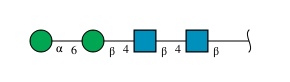
alpha-D-mannopyranose-(1-6)-beta-D-mannopyranose-(1-4)-2-acetamido-2-deoxy-beta-D-glucopyranose-(1-4)-2-acetamido-2-deoxy-beta-D-glucopyranoseC [auth S]4N-Glycosylation
Glycosylation Resources
GlyTouCan: G22573RC
GlyCosmos: G22573RC
GlyGen: G22573RC

Experimental Data & Validation

Experimental Data

  • Method: X-RAY DIFFRACTION
  • Resolution: 1.38 Å
  • R-Value Free:  0.217 (Depositor), 0.222 (DCC) 
  • R-Value Work:  0.195 (Depositor), 0.200 (DCC) 
  • R-Value Observed: 0.197 (Depositor) 
Space Group: P 21 21 21
Unit Cell:
Length ( Å )Angle ( ˚ )
a = 62.93α = 90
b = 80.882β = 90
c = 87.469γ = 90
Software Package:
Software NamePurpose
REFMACrefinement
DIALSdata reduction
Aimlessdata scaling
PHASERphasing

Structure Validation

View Full Validation Report


Entry History 

& Funding Information

Deposition Data


Funding OrganizationLocationGrant Number
National Institutes of Health/National Cancer Institute (NIH/NCI)United StatesP30 CA015704-40
Norwegian Research CouncilNorway245828

Revision History  (Full details and data files)

  • Version 1.0: 2024-09-25
    Type: Initial release
  • Version 1.1: 2025-02-05
    Changes: Database references, Structure summary
  • Version 1.2: 2025-10-22
    Changes: Database references