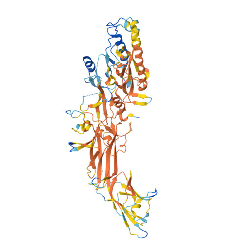
9B98

Crystal structure of the human PAD2 protein bound to small molecule


Experimental Data Snapshot

  • Method: X-RAY DIFFRACTION
  • Resolution: 2.00 Å
  • R-Value Free: 0.258 
  • R-Value Work: 0.186 

Starting Model: experimental
View more details

wwPDB Validation   3D Report Full Report


Ligand Structure Quality Assessment 


This is version 1.1 of the entry. See complete history


Literature

Discovery, Characterization, and Structure of a Cell Active PAD2 Inhibitor Acting through a Novel Allosteric Mechanism.

Byrnes, L.J.Choi, W.Y.Balbo, P.Banker, M.E.Chang, J.Chen, S.Cheng, X.Cong, Y.Culp, J.Di, H.Griffor, M.Hall, J.Meng, X.Morgan, B.Mousseau, J.J.Nicki, J.O'Connell, T.Ramsey, S.Shaginian, A.Shanker, S.Trujillo, J.Wan, J.Vincent, F.Wright, S.W.Vajdos, F.

(2024) ACS Chem Biol 19: 2186-2197

  • DOI: https://doi.org/10.1021/acschembio.4c00397
  • Primary Citation of Related Structures:  
    9B96, 9B97, 9B98

  • PubMed Abstract: 

    Peptidyl arginine deiminases (PADs) are important enzymes in many diseases, especially those involving inflammation and autoimmunity. Despite many years of effort, developing isoform-specific inhibitors has been a challenge. We describe herein the discovery of a potent, noncovalent PAD2 inhibitor, with selectivity over PAD3 and PAD4, from a DNA-encoded library. The biochemical and biophysical characterization of this inhibitor and two noninhibitory binders indicated a novel, Ca 2+ competitive mechanism of inhibition. This was confirmed via X-ray crystallographic analysis. Finally, we demonstrate that this inhibitor selectively inhibits PAD2 in a cellular context.


  • Organizational Affiliation

    Pfizer Worldwide Research and Development, Eastern Pt. Rd, Groton, Connecticut 06340-5146, United States.


Macromolecules
Find similar proteins by:  (by identity cutoff)  |  3D Structure
Entity ID: 1
MoleculeChains Sequence LengthOrganismDetailsImage
Protein-arginine deiminase type-2690Homo sapiensMutation(s): 0 
Gene Names: PADI2KIAA0994PAD2PDI2
EC: 3.5.3.15
UniProt & NIH Common Fund Data Resources
Find proteins for Q9Y2J8 (Homo sapiens)
Explore Q9Y2J8 
Go to UniProtKB:  Q9Y2J8
PHAROS:  Q9Y2J8
GTEx:  ENSG00000117115 
Entity Groups  
Sequence Clusters30% Identity50% Identity70% Identity90% Identity95% Identity100% Identity
UniProt GroupQ9Y2J8
Sequence Annotations
Expand
  • Reference Sequence
Small Molecules
Ligands 3 Unique
IDChains Name / Formula / InChI Key2D Diagram3D Interactions
A1AJC (Subject of Investigation/LOI)
Query on A1AJC

Download Ideal Coordinates CCD File 
I [auth A](5P)-N,N-diethyl-2-fluoro-5-(2-[({1-[2-(methylamino)-2-oxoethyl]cyclohexyl}methyl)amino]-6-{methyl[1-(2-methyl-1-phenyl-1H-1,3-benzimidazole-5-carbonyl)piperidin-4-yl]amino}pyrimidin-4-yl)benzamide
C46 H56 F N9 O3
HMDXTOLTWKZGHP-UHFFFAOYSA-N
ACT
Query on ACT

Download Ideal Coordinates CCD File 
B [auth A],
C [auth A]
ACETATE ION
C2 H3 O2
QTBSBXVTEAMEQO-UHFFFAOYSA-M
CA
Query on CA

Download Ideal Coordinates CCD File 
D [auth A],
E [auth A],
F [auth A],
G [auth A],
H [auth A]
CALCIUM ION
Ca
BHPQYMZQTOCNFJ-UHFFFAOYSA-N
Experimental Data & Validation

Experimental Data

  • Method: X-RAY DIFFRACTION
  • Resolution: 2.00 Å
  • R-Value Free: 0.258 
  • R-Value Work: 0.186 
  • Space Group: C 1 2 1
Unit Cell:
Length ( Å )Angle ( ˚ )
a = 201.71α = 90
b = 52.78β = 105.709
c = 76.59γ = 90
Software Package:
Software NamePurpose
REFMACrefinement
autoPROCdata reduction
autoPROCdata scaling
PHASERphasing

Structure Validation

View Full Validation Report


Ligand Structure Quality Assessment 


Entry History & Funding Information

Deposition Data


Funding OrganizationLocationGrant Number
Other private--

Revision History  (Full details and data files)

  • Version 1.0: 2024-10-09
    Type: Initial release
  • Version 1.1: 2024-10-30
    Changes: Database references