9B69

GluA2 flip Q in complex with TARPgamma2 at pH8, class12, structure of NTD

  • Classification: TRANSPORT PROTEIN
  • Organism(s): Rattus norvegicus
  • Expression System: Homo sapiens
  • Mutation(s): No 

  • Deposited: 2024-03-23 Released: 2024-07-31 
  • Deposition Author(s): Nakagawa, T., Greger, I.H.
  • Funding Organization(s): National Institutes of Health/National Institute of Mental Health (NIH/NIMH), UK Research and Innovation (UKRI), Biotechnology and Biological Sciences Research Council (BBSRC), Wellcome Trust

Experimental Data Snapshot

  • Method: ELECTRON MICROSCOPY
  • Resolution: 3.69 Å
  • Aggregation State: PARTICLE 
  • Reconstruction Method: SINGLE PARTICLE 

Starting Models: experimental
View more details

wwPDB Validation   3D Report Full Report


This is version 1.2 of the entry. See complete history


Literature

Proton-triggered rearrangement of the AMPA receptor N-terminal domains impacts receptor kinetics and synaptic localization.

Ivica, J.Kejzar, N.Ho, H.Stockwell, I.Kuchtiak, V.Scrutton, A.M.Nakagawa, T.Greger, I.H.

(2024) Nat Struct Mol Biol 

  • DOI: https://doi.org/10.1038/s41594-024-01369-5
  • Primary Citation of Related Structures:  
    9B5Z, 9B60, 9B61, 9B63, 9B64, 9B67, 9B68, 9B69, 9B6A

  • PubMed Abstract: 

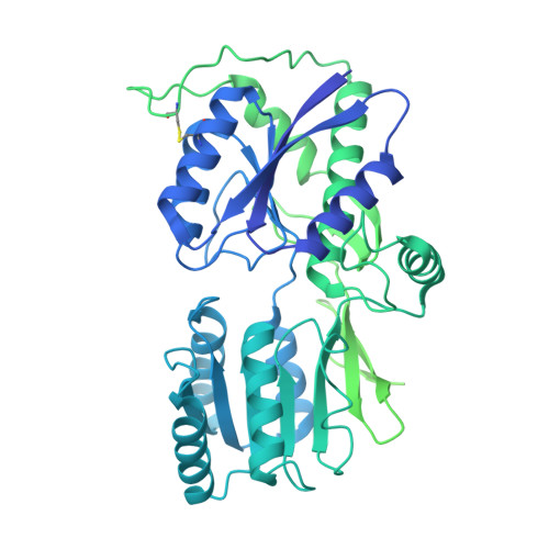
    AMPA glutamate receptors (AMPARs) are ion channel tetramers that mediate the majority of fast excitatory synaptic transmission. They are composed of four subunits (GluA1-GluA4); the GluA2 subunit dominates AMPAR function throughout the forebrain. Its extracellular N-terminal domain (NTD) determines receptor localization at the synapse, ensuring reliable synaptic transmission and plasticity. This synaptic anchoring function requires a compact NTD tier, stabilized by a GluA2-specific NTD interface. Here we show that low pH conditions, which accompany synaptic activity, rupture this interface. All-atom molecular dynamics simulations reveal that protonation of an interfacial histidine residue (H208) centrally contributes to NTD rearrangement. Moreover, in stark contrast to their canonical compact arrangement at neutral pH, GluA2 cryo-electron microscopy structures exhibit a wide spectrum of NTD conformations under acidic conditions. We show that the consequences of this pH-dependent conformational control are twofold: rupture of the NTD tier slows recovery from desensitized states and increases receptor mobility at mouse hippocampal synapses. Therefore, a proton-triggered NTD switch will shape both AMPAR location and kinetics, thereby impacting synaptic signal transmission.


  • Organizational Affiliation

    Neurobiology Division, Medical Research Council (MRC) Laboratory of Molecular Biology, Cambridge, UK.


Macromolecules
Find similar proteins by:  (by identity cutoff)  |  3D Structure
Entity ID: 1
MoleculeChains Sequence LengthOrganismDetailsImage
Isoform Flip of Glutamate receptor 2A [auth C],
B [auth A],
C [auth B],
D
889Rattus norvegicusMutation(s): 0 
Gene Names: Gria2Glur2
UniProt
Find proteins for P19491 (Rattus norvegicus)
Explore P19491 
Go to UniProtKB:  P19491
Entity Groups  
Sequence Clusters30% Identity50% Identity70% Identity90% Identity95% Identity100% Identity
UniProt GroupP19491
Glycosylation
Glycosylation Sites: 1Go to GlyGen: P19491-1
Sequence Annotations
Expand
  • Reference Sequence
Oligosaccharides

Help

Entity ID: 2
MoleculeChains Length2D Diagram Glycosylation3D Interactions
2-acetamido-2-deoxy-beta-D-glucopyranose-(1-4)-2-acetamido-2-deoxy-beta-D-glucopyranose
E, H
2N-Glycosylation
Glycosylation Resources
GlyTouCan:  G42666HT
GlyCosmos:  G42666HT
GlyGen:  G42666HT
Entity ID: 3
MoleculeChains Length2D Diagram Glycosylation3D Interactions
beta-D-mannopyranose-(1-4)-2-acetamido-2-deoxy-beta-D-glucopyranose-(1-4)-2-acetamido-2-deoxy-beta-D-glucopyranose
F, G
3N-Glycosylation
Glycosylation Resources
GlyTouCan:  G15407YE
GlyCosmos:  G15407YE
GlyGen:  G15407YE
Small Molecules
Ligands 1 Unique
IDChains Name / Formula / InChI Key2D Diagram3D Interactions
NAG (Subject of Investigation/LOI)
Query on NAG

Download Ideal Coordinates CCD File 
I [auth B]2-acetamido-2-deoxy-beta-D-glucopyranose
C8 H15 N O6
OVRNDRQMDRJTHS-FMDGEEDCSA-N
Experimental Data & Validation

Experimental Data

  • Method: ELECTRON MICROSCOPY
  • Resolution: 3.69 Å
  • Aggregation State: PARTICLE 
  • Reconstruction Method: SINGLE PARTICLE 
EM Software:
TaskSoftware PackageVersion
MODEL REFINEMENTPHENIX1.20.1_4487:
RECONSTRUCTIONRELION4

Structure Validation

View Full Validation Report


Entry History & Funding Information

Deposition Data


Funding OrganizationLocationGrant Number
National Institutes of Health/National Institute of Mental Health (NIH/NIMH)United StatesR01MH123474
UK Research and Innovation (UKRI)United KingdomMC_U105174197
Biotechnology and Biological Sciences Research Council (BBSRC)United KingdomBB/N002113/1
Wellcome TrustUnited Kingdom223194/Z/21/Z

Revision History  (Full details and data files)

  • Version 1.0: 2024-07-31
    Type: Initial release
  • Version 1.1: 2024-08-21
    Changes: Data collection, Database references
  • Version 1.2: 2024-09-04
    Changes: Data collection, Database references