9B1D

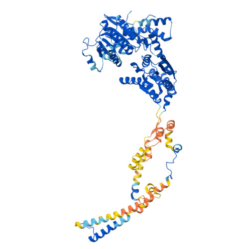
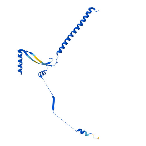
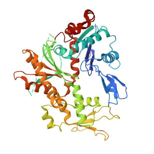
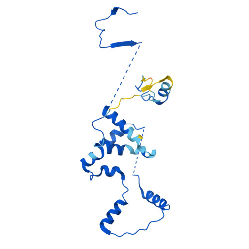
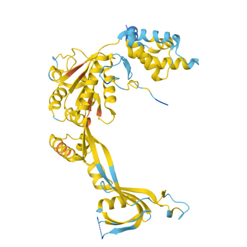
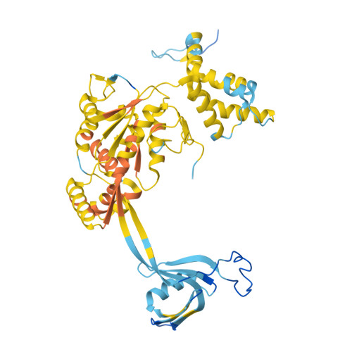
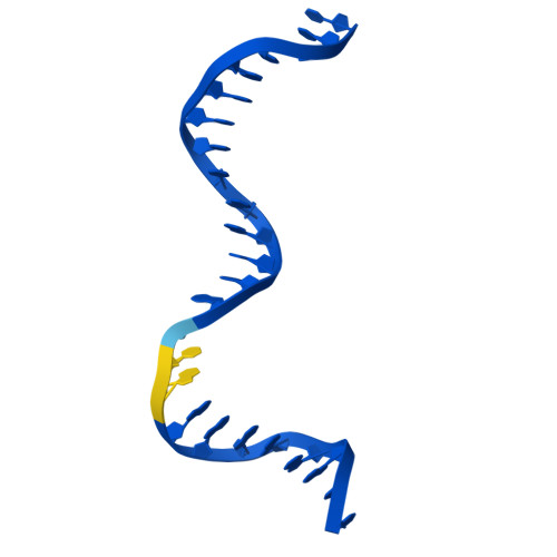
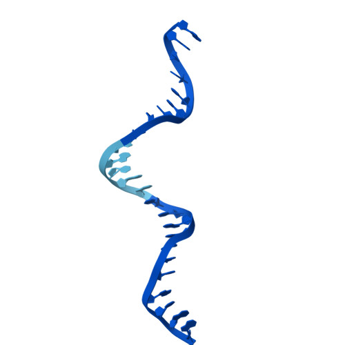
Cryo-EM structure of native SWR1 bound to DNA (composite structure)


Experimental Data Snapshot

  • Method: ELECTRON MICROSCOPY
  • Resolution: 3.30 Å
  • Aggregation State: PARTICLE 
  • Reconstruction Method: SINGLE PARTICLE 

Starting Models: experimental, in silico
View more details

wwPDB Validation   3D Report Full Report


This is version 1.1 of the entry. See complete history


Literature

Molecular basis of global promoter sensing and nucleosome capture by the SWR1 chromatin remodeler.

Louder, R.K.Park, G.Ye, Z.Cha, J.S.Gardner, A.M.Lei, Q.Ranjan, A.Hollmuller, E.Stengel, F.Pugh, B.F.Wu, C.

(2024) Cell 

  • DOI: https://doi.org/10.1016/j.cell.2024.09.007
  • Primary Citation of Related Structures:  
    9B1D, 9B1E

  • PubMed Abstract: 

    The SWR1 chromatin remodeling complex is recruited to +1 nucleosomes downstream of transcription start sites of eukaryotic promoters, where it exchanges histone H2A for the specialized variant H2A.Z. Here, we use cryoelectron microscopy (cryo-EM) to resolve the structural basis of the SWR1 interaction with free DNA, revealing a distinct open conformation of the Swr1 ATPase that enables sliding from accessible DNA to nucleosomes. A complete structural model of the SWR1-nucleosome complex illustrates critical roles for Swc2 and Swc3 subunits in oriented nucleosome engagement by SWR1. Moreover, an extended DNA-binding α helix within the Swc3 subunit enables sensing of nucleosome linker length and is essential for SWR1-promoter-specific recruitment and activity. The previously unresolved N-SWR1 subcomplex forms a flexible extended structure, enabling multivalent recognition of acetylated histone tails by reader domains to further direct SWR1 toward the +1 nucleosome. Altogether, our findings provide a generalizable mechanism for promoter-specific targeting of chromatin and transcription complexes.


  • Organizational Affiliation

    Department of Biology, Johns Hopkins University, Baltimore, MD, USA. Electronic address: rklouder@jhu.edu.


Macromolecules

Find similar proteins by:  (by identity cutoff)  |  3D Structure
Entity ID: 1
MoleculeChains Sequence LengthOrganismDetailsImage
Helicase SWR11,544Saccharomyces cerevisiae W303Mutation(s): 0 
EC: 3.6.4.12
Entity Groups  
Sequence Clusters30% Identity50% Identity70% Identity90% Identity95% Identity100% Identity
Sequence Annotations
Expand
  • Reference Sequence
Find similar proteins by:  (by identity cutoff)  |  3D Structure
Entity ID: 2
MoleculeChains Sequence LengthOrganismDetailsImage
Vacuolar protein sorting-associated protein 72795Saccharomyces cerevisiae W303Mutation(s): 0 
UniProt
Find proteins for Q03388 (Saccharomyces cerevisiae (strain ATCC 204508 / S288c))
Explore Q03388 
Go to UniProtKB:  Q03388
Entity Groups  
Sequence Clusters30% Identity50% Identity70% Identity90% Identity95% Identity100% Identity
UniProt GroupQ03388
Sequence Annotations
Expand
  • Reference Sequence
Find similar proteins by:  (by identity cutoff)  |  3D Structure
Entity ID: 3
MoleculeChains Sequence LengthOrganismDetailsImage
Actin-like protein ARP6438Saccharomyces cerevisiae W303Mutation(s): 0 
UniProt
Find proteins for Q12509 (Saccharomyces cerevisiae (strain ATCC 204508 / S288c))
Explore Q12509 
Go to UniProtKB:  Q12509
Entity Groups  
Sequence Clusters30% Identity50% Identity70% Identity90% Identity95% Identity100% Identity
UniProt GroupQ12509
Sequence Annotations
Expand
  • Reference Sequence
Find similar proteins by:  (by identity cutoff)  |  3D Structure
Entity ID: 4
MoleculeChains Sequence LengthOrganismDetailsImage
Vacuolar protein sorting-associated protein 71280Saccharomyces cerevisiae W303Mutation(s): 0 
UniProt
Find proteins for Q03433 (Saccharomyces cerevisiae (strain ATCC 204508 / S288c))
Explore Q03433 
Go to UniProtKB:  Q03433
Entity Groups  
Sequence Clusters30% Identity50% Identity70% Identity90% Identity95% Identity100% Identity
UniProt GroupQ03433
Sequence Annotations
Expand
  • Reference Sequence
Find similar proteins by:  (by identity cutoff)  |  3D Structure
Entity ID: 5
MoleculeChains Sequence LengthOrganismDetailsImage
RuvB-like protein 1
E, G, I
837Saccharomyces cerevisiae W303Mutation(s): 0 
EC: 3.6.4.12
Entity Groups  
Sequence Clusters30% Identity50% Identity70% Identity90% Identity95% Identity100% Identity
Sequence Annotations
Expand
  • Reference Sequence
Find similar proteins by:  (by identity cutoff)  |  3D Structure
Entity ID: 6
MoleculeChains Sequence LengthOrganismDetailsImage
RuvB-like protein 2
F, H, J
471Saccharomyces cerevisiae W303Mutation(s): 0 
EC: 3.6.4.12
UniProt
Find proteins for Q12464 (Saccharomyces cerevisiae (strain ATCC 204508 / S288c))
Explore Q12464 
Go to UniProtKB:  Q12464
Entity Groups  
Sequence Clusters30% Identity50% Identity70% Identity90% Identity95% Identity100% Identity
UniProt GroupQ12464
Sequence Annotations
Expand
  • Reference Sequence
Find similar nucleic acids by:  (by identity cutoff)  |  3D Structure
Entity ID: 7
MoleculeChains LengthOrganismImage
DNA (147-MER)K [auth Y]147synthetic construct
Sequence Annotations
Expand
  • Reference Sequence
Find similar nucleic acids by:  (by identity cutoff)  |  3D Structure
Entity ID: 8
MoleculeChains LengthOrganismImage
DNA (147-MER)L [auth Z]147synthetic construct
Sequence Annotations
Expand
  • Reference Sequence
Small Molecules
Ligands 4 Unique
IDChains Name / Formula / InChI Key2D Diagram3D Interactions
AGS
Query on AGS

Download Ideal Coordinates CCD File 
M [auth A],
O [auth C],
S [auth E],
X [auth G]
PHOSPHOTHIOPHOSPHORIC ACID-ADENYLATE ESTER
C10 H16 N5 O12 P3 S
NLTUCYMLOPLUHL-KQYNXXCUSA-N
ADP
Query on ADP

Download Ideal Coordinates CCD File 
AA [auth I],
CA [auth J],
V [auth F],
Z [auth H]
ADENOSINE-5'-DIPHOSPHATE
C10 H15 N5 O10 P2
XTWYTFMLZFPYCI-KQYNXXCUSA-N
ZN
Query on ZN

Download Ideal Coordinates CCD File 
Q [auth D],
R [auth D]
ZINC ION
Zn
PTFCDOFLOPIGGS-UHFFFAOYSA-N
MG
Query on MG

Download Ideal Coordinates CCD File 
BA [auth I]
DA [auth J]
N [auth A]
P [auth C]
T [auth E]
BA [auth I],
DA [auth J],
N [auth A],
P [auth C],
T [auth E],
U [auth F],
W [auth G],
Y [auth H]
MAGNESIUM ION
Mg
JLVVSXFLKOJNIY-UHFFFAOYSA-N
Experimental Data & Validation

Experimental Data

  • Method: ELECTRON MICROSCOPY
  • Resolution: 3.30 Å
  • Aggregation State: PARTICLE 
  • Reconstruction Method: SINGLE PARTICLE 
EM Software:
TaskSoftware PackageVersion
RECONSTRUCTIONRELION3.1.3
MODEL REFINEMENTPHENIX1.21_5207

Structure Validation

View Full Validation Report


Entry History & Funding Information

Deposition Data


Funding OrganizationLocationGrant Number
National Institutes of Health/National Institute of General Medical Sciences (NIH/NIGMS)United StatesR35GM149291
National Institutes of Health/National Institute of General Medical Sciences (NIH/NIGMS)United StatesR01GM125831
National Institutes of Health/National Institute of General Medical Sciences (NIH/NIGMS)United StatesF32GM133151

Revision History  (Full details and data files)

  • Version 1.0: 2024-10-09
    Type: Initial release
  • Version 1.1: 2024-10-16
    Changes: Data collection, Database references