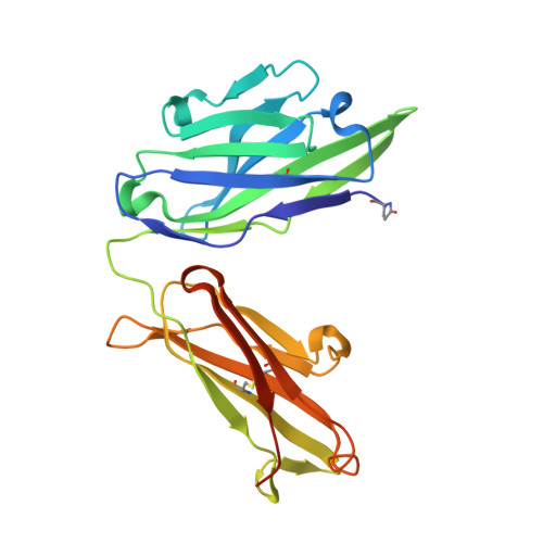
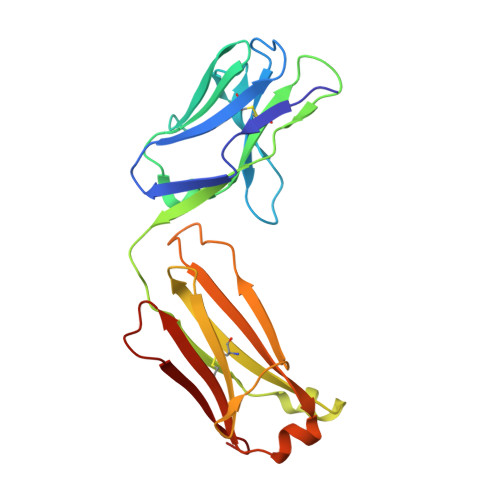
Structure-Based Engineering of Monoclonal Antibodies for Improved Binding to Counteract the Effects of Fentanyl and Carfentanil.
Rodarte, J., Baehr, C., Hicks, D., McGovern, M., Zhang, Y., Silva-Ortiz, P., Hannon, B., Duddu, S., Pancera, M., Pravetoni, M.(2024) ACS Omega 9: 42506-42519
- PubMed: 39431098 Search on PubMedSearch on PubMed Central
- DOI: https://doi.org/10.1021/acsomega.4c06617
- Primary Citation Related Structures:
9AXN, 9AXO, 9AXP, 9AXQ, 9AXR, 9AXS - PubMed Abstract:
The opioid overdose epidemic is a growing and evolving public health crisis fueled by the widespread presence of fentanyl and fentanyl analogues (F/FAs) in both street mixtures and counterfeit pills. To expand current treatment options, drug-targeting monoclonal antibodies (mAbs) offer a viable therapeutic for both pre- and postexposure clinical scenarios. This study reports the isolation, in vitro characterization, and in vivo efficacy of two murine mAb families targeting fentanyl, carfentanil, or both. Because humanization of the mAbs by CDR grafting negatively impacted affinity for both fentanyl and carfentanil, crystal structures of mAbs in complex with fentanyl or carfentanil were analyzed to identify key residues involved in ligand binding in murine versus humanized structures, and site-directed mutagenesis was used to verify their functional importance. The structural analysis identified a framework residue, Tyr36, present in the murine germline sequence of two mAbs, which was critical for binding to fentanyl and carfentanil. These studies emphasize the importance of structural considerations in mAb engineering to optimize mAbs targeting small molecules including opioids and other drugs of public health interest.
- Vaccine and Infectious Disease Division, Fred Hutchinson Cancer Center, Seattle, Washington 98109, United States.
Organizational Affiliation: