9AXF | pdb_00009axf

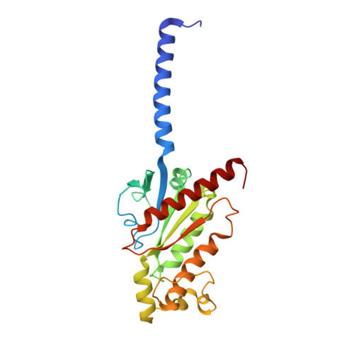
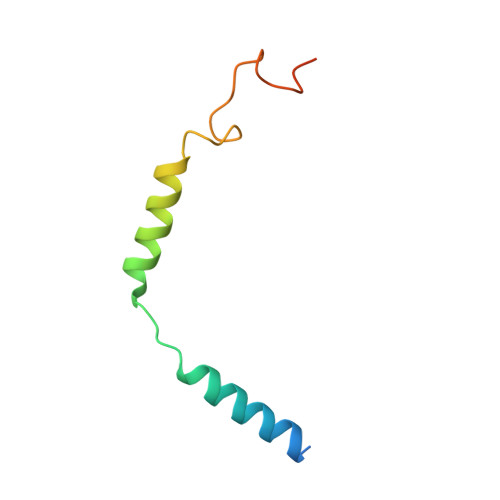
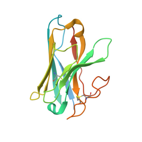
Structure of human calcium-sensing receptor in complex with chimeric Gq (miniGisq) protein in detergent


Experimental Data Snapshot

  • Method: ELECTRON MICROSCOPY
  • Resolution: 3.50 Å
  • Aggregation State: PARTICLE 
  • Reconstruction Method: SINGLE PARTICLE 

Starting Models: experimental
View more details

wwPDB Validation 3D Report Full Report

Validation slider image for 9AXF

This is version 1.3 of the entry. See complete history. 

Literature

Promiscuous G-protein activation by the calcium-sensing receptor.

Zuo, H., Park, J., Frangaj, A., Ye, J., Lu, G., Manning, J.J., Asher, W.B., Lu, Z., Hu, G.B., Wang, L., Mendez, J., Eng, E., Zhang, Z., Lin, X., Grassucci, R., Hendrickson, W.A., Clarke, O.B., Javitch, J.A., Conigrave, A.D., Fan, Q.R.

(2024) Nature 629: 481-488

  • DOI: https://doi.org/10.1038/s41586-024-07331-1
  • Primary Citation Related Structures: 
    9ASB, 9AVG, 9AVL, 9AXF, 9AYF

  • PubMed Abstract: 

    The human calcium-sensing receptor (CaSR) detects fluctuations in the extracellular Ca 2+ concentration and maintains Ca 2+ homeostasis 1,2 . It also mediates diverse cellular processes not associated with Ca 2+ balance 3-5 . The functional pleiotropy of CaSR arises in part from its ability to signal through several G-protein subtypes 6 . We determined structures of CaSR in complex with G proteins from three different subfamilies: G q , G i and G s . We found that the homodimeric CaSR of each complex couples to a single G protein through a common mode. This involves the C-terminal helix of each Gα subunit binding to a shallow pocket that is formed in one CaSR subunit by all three intracellular loops (ICL1-ICL3), an extended transmembrane helix 3 and an ordered C-terminal region. G-protein binding expands the transmembrane dimer interface, which is further stabilized by phospholipid. The restraint imposed by the receptor dimer, in combination with ICL2, enables G-protein activation by facilitating conformational transition of Gα. We identified a single Gα residue that determines G q and G s versus G i selectivity. The length and flexibility of ICL2 allows CaSR to bind all three Gα subtypes, thereby conferring capacity for promiscuous G-protein coupling.


  • Organizational Affiliation: 
    • Department of Molecular Pharmacology and Therapeutics, Columbia University, New York, NY, USA.

Macromolecule Content 

  • Total Structure Weight: 340.56 kDa 
  • Atom Count: 21,342 
  • Modeled Residue Count: 2,605 
  • Deposited Residue Count: 2,944 
  • Unique protein chains: 6

Macromolecules

Find similar proteins by:|  3D Structure
Entity ID: 1
MoleculeChains  Sequence LengthOrganismDetailsImage
Extracellular calcium-sensing receptorA [auth R],
B [auth Q]
911Homo sapiensMutation(s): 0 
Gene Names: CASR, GPRC2A, PCAR1
Membrane Entity: Yes 
UniProt & NIH Common Fund Data Resources
Find proteins for P41180 (Homo sapiens)
Explore P41180 
Go to UniProtKB:  P41180
PHAROS:  P41180
GTEx:  ENSG00000036828 
Entity Groups
Sequence Clusters30% Identity50% Identity70% Identity90% Identity95% Identity100% Identity
UniProt GroupP41180
Glycosylation
Glycosylation Sites: 6Go to GlyGen: P41180-1
Sequence Annotations
Expand
Reference Sequence
Find similar proteins by:|  3D Structure
Entity ID: 2
MoleculeChains  Sequence LengthOrganismDetailsImage
Guanine nucleotide-binding protein G(i) subunit alpha-1,Adenylate cyclase-stimulating G alpha proteinC [auth A]246Homo sapiensMutation(s): 28 
Gene Names: GNAI1, GNAS
EC: 3.6.5
Membrane Entity: Yes 
UniProt & NIH Common Fund Data Resources
Find proteins for P63092 (Homo sapiens)
Explore P63092 
Go to UniProtKB:  P63092
PHAROS:  P63092
GTEx:  ENSG00000087460 
Entity Groups
Sequence Clusters30% Identity50% Identity70% Identity90% Identity95% Identity100% Identity
UniProt GroupP63092
Sequence Annotations
Expand
Reference Sequence
Find similar proteins by:|  3D Structure
Entity ID: 3
MoleculeChains  Sequence LengthOrganismDetailsImage
Guanine nucleotide-binding protein G(I)/G(S)/G(T) subunit beta-1D [auth B]348Homo sapiensMutation(s): 0 
Gene Names: GNB1
UniProt & NIH Common Fund Data Resources
Find proteins for P62873 (Homo sapiens)
Explore P62873 
Go to UniProtKB:  P62873
PHAROS:  P62873
GTEx:  ENSG00000078369 
Entity Groups
Sequence Clusters30% Identity50% Identity70% Identity90% Identity95% Identity100% Identity
UniProt GroupP62873
Sequence Annotations
Expand
Reference Sequence
Find similar proteins by:|  3D Structure
Entity ID: 4
MoleculeChains  Sequence LengthOrganismDetailsImage
Guanine nucleotide-binding protein G(I)/G(S)/G(O) subunit gamma-2E [auth G]71Homo sapiensMutation(s): 0 
Gene Names: GNG2
Membrane Entity: Yes 
UniProt & NIH Common Fund Data Resources
Find proteins for P59768 (Homo sapiens)
Explore P59768 
Go to UniProtKB:  P59768
PHAROS:  P59768
GTEx:  ENSG00000186469 
Entity Groups
Sequence Clusters30% Identity50% Identity70% Identity90% Identity95% Identity100% Identity
UniProt GroupP59768
Sequence Annotations
Expand
Reference Sequence
Find similar proteins by:|  3D Structure
Entity ID: 5
MoleculeChains  Sequence LengthOrganismDetailsImage
Single-chain antibody fragment scFv16F [auth H]297Mus musculusMutation(s): 0 
Entity Groups
Sequence Clusters30% Identity50% Identity70% Identity90% Identity95% Identity100% Identity
Sequence Annotations
Expand
Reference Sequence
Find similar proteins by:|  3D Structure
Entity ID: 6
MoleculeChains  Sequence LengthOrganismDetailsImage
Nanobody Nb-35G [auth N]160Lama glamaMutation(s): 0 
Entity Groups
Sequence Clusters30% Identity50% Identity70% Identity90% Identity95% Identity100% Identity
Sequence Annotations
Expand
Reference Sequence

Oligosaccharides

Help  
Entity ID: 7
MoleculeChains Length2D Diagram GlycosylationD Interactions
2-acetamido-2-deoxy-beta-D-glucopyranose-(1-4)-2-acetamido-2-deoxy-beta-D-glucopyranoseH [auth C],
I [auth D],
J [auth E],
K [auth F]
2N-Glycosylation
Glycosylation Resources
GlyTouCan: G42666HT
GlyCosmos: G42666HT
GlyGen: G42666HT

Small Molecules

Ligands 8 Unique
IDChains Name / Formula / InChI Key2D Diagram3D Interactions
AV0

Query on AV0


Download:Ideal Coordinates CCD File
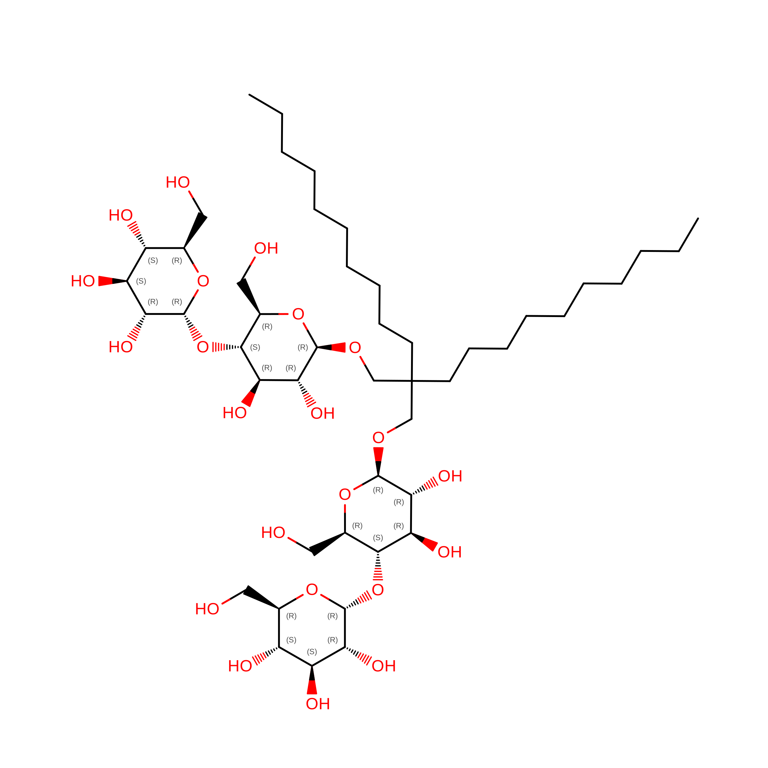
V [auth R]Lauryl Maltose Neopentyl Glycol
C47 H88 O22
MADJBYLAYPCCOO-VWHTXWAPSA-N
Y01

Query on Y01


Download:Ideal Coordinates CCD File
NA [auth Q],
W [auth R],
X [auth R],
Y [auth R]
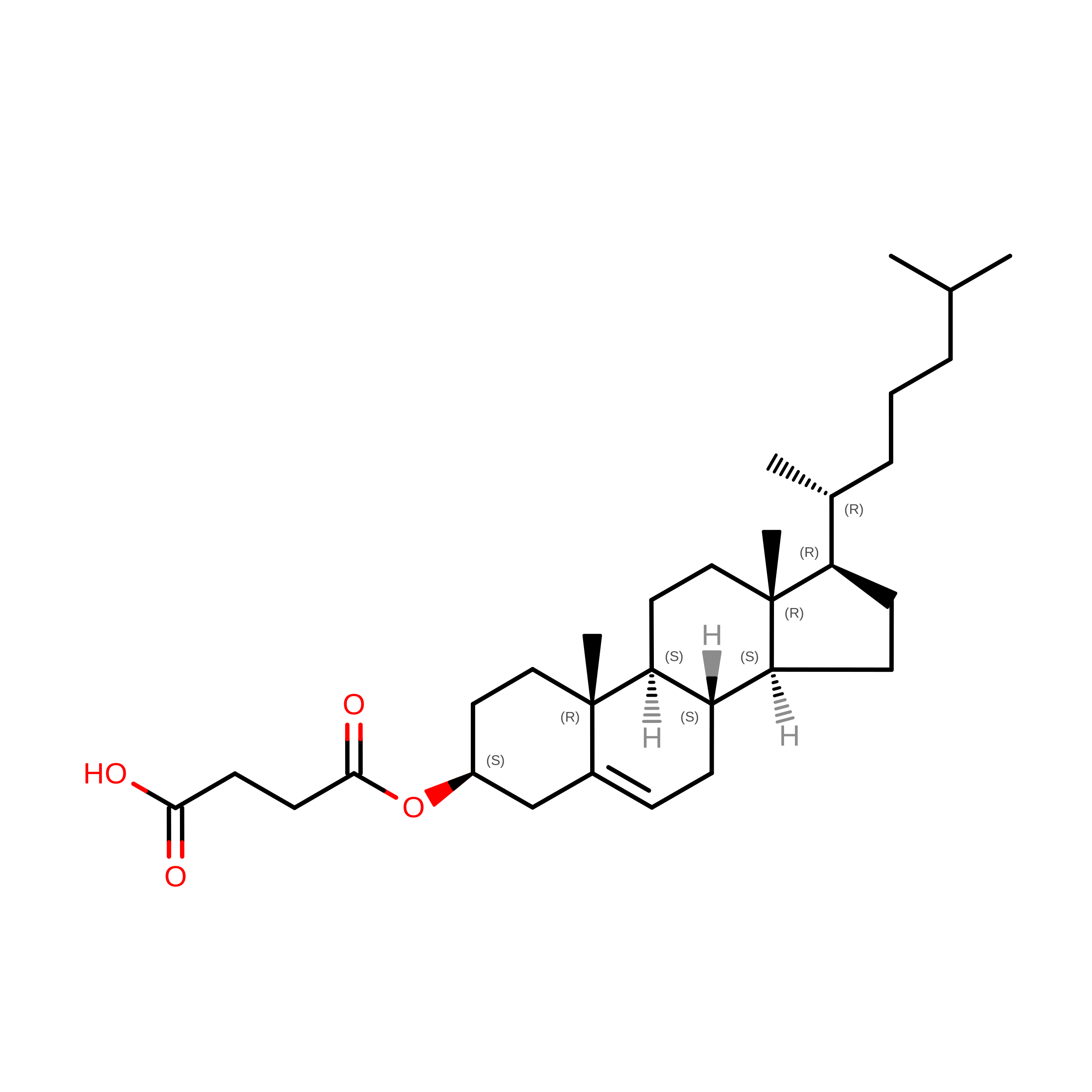
CHOLESTEROL HEMISUCCINATE
C31 H50 O4
WLNARFZDISHUGS-MIXBDBMTSA-N
CLR

Query on CLR


Download:Ideal Coordinates CCD File
AA [auth R]
BA [auth R]
OA [auth Q]
PA [auth Q]
QA [auth Q]
AA [auth R],
BA [auth R],
OA [auth Q],
PA [auth Q],
QA [auth Q],
RA [auth Q],
SA [auth Q],
Z [auth R]
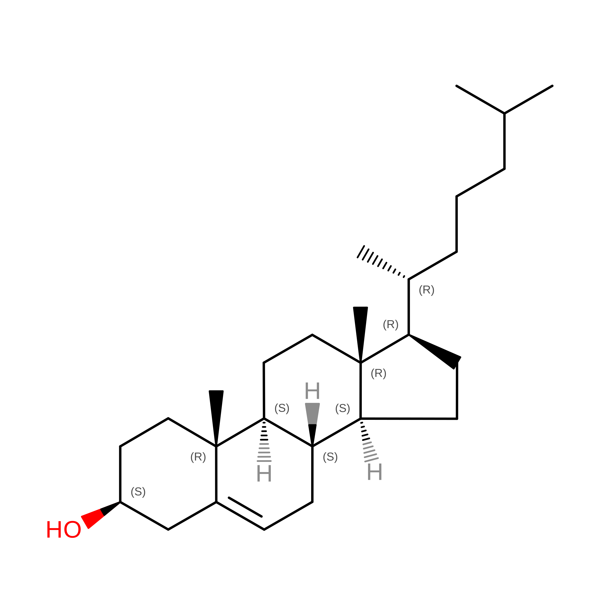
CHOLESTEROL
C27 H46 O
HVYWMOMLDIMFJA-DPAQBDIFSA-N
9IG

Query on 9IG


Download:Ideal Coordinates CCD File
MA [auth Q],
U [auth R]
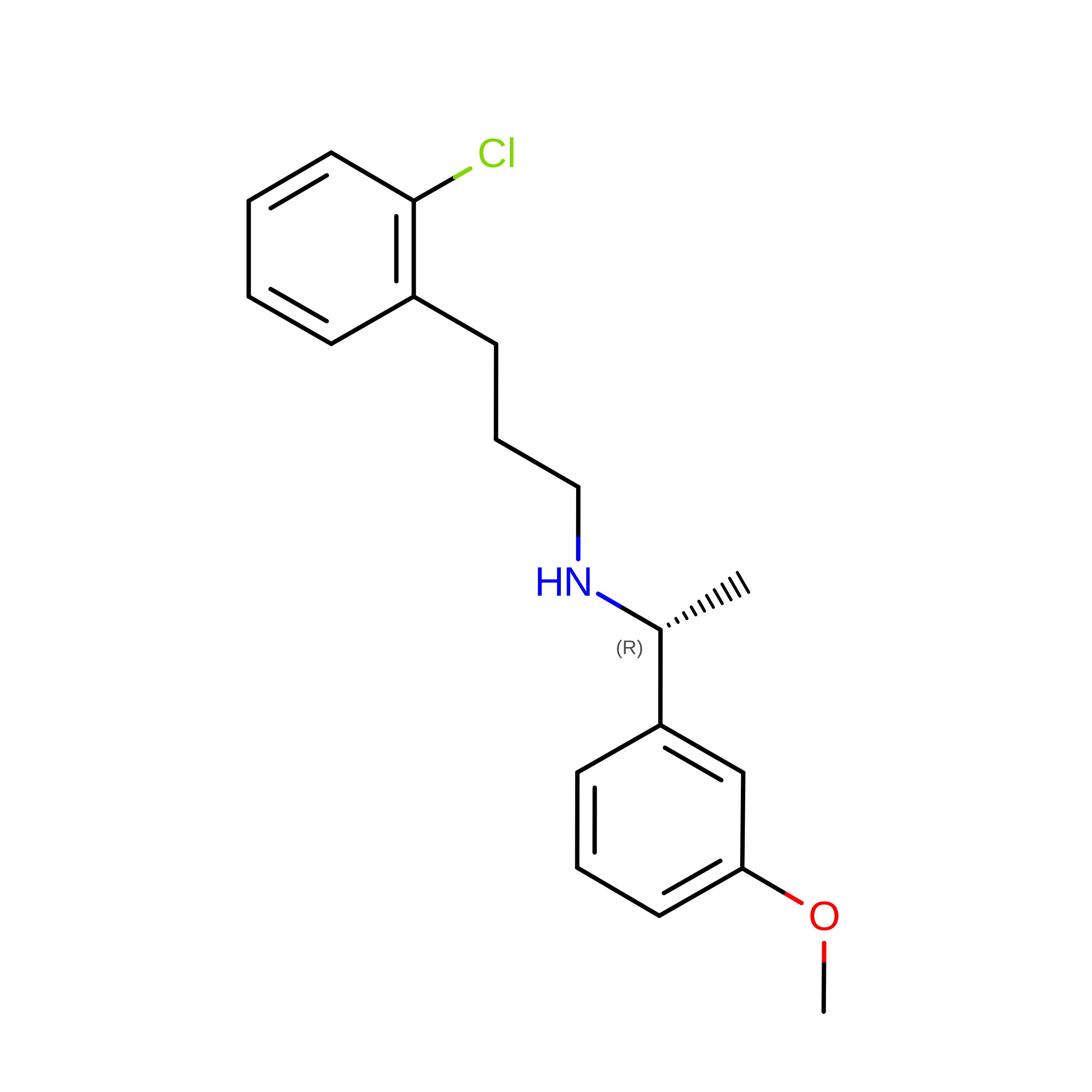
3-(2-chlorophenyl)-N-[(1R)-1-(3-methoxyphenyl)ethyl]propan-1-amine
C18 H22 Cl N O
ZVQUCWXZCKWZBP-CQSZACIVSA-N
NAG

Query on NAG


Download:Ideal Coordinates CCD File
CA [auth Q]
DA [auth Q]
EA [auth Q]
FA [auth Q]
L [auth R]
CA [auth Q],
DA [auth Q],
EA [auth Q],
FA [auth Q],
L [auth R],
M [auth R],
N [auth R],
O [auth R]
2-acetamido-2-deoxy-beta-D-glucopyranose
C8 H15 N O6
OVRNDRQMDRJTHS-FMDGEEDCSA-N
TCR

Query on TCR


Download:Ideal Coordinates CCD File
GA [auth Q],
P [auth R]
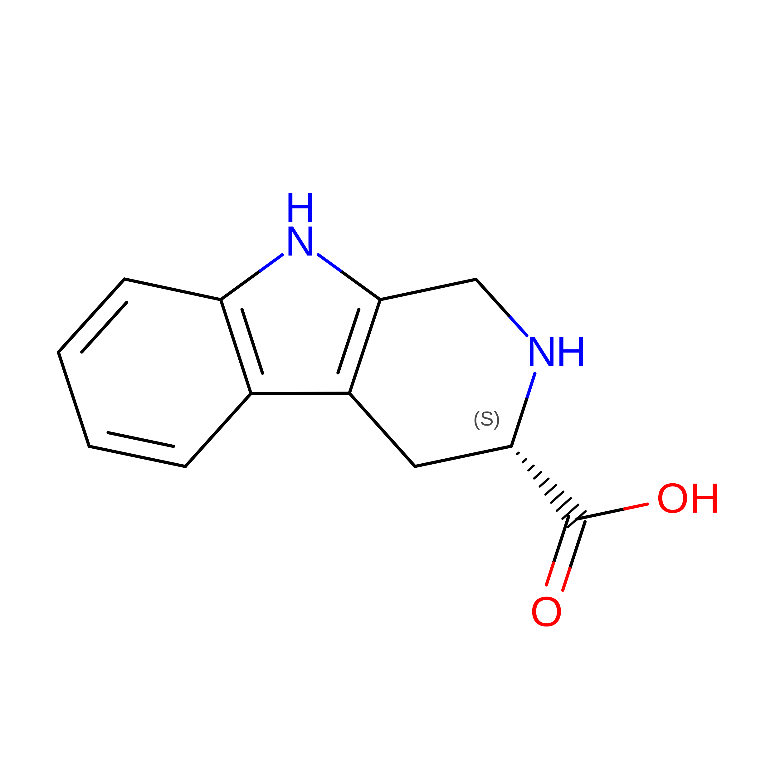
CYCLOMETHYLTRYPTOPHAN
C12 H12 N2 O2
FSNCEEGOMTYXKY-JTQLQIEISA-N
PO4

Query on PO4


Download:Ideal Coordinates CCD File
HA [auth Q],
Q [auth R]
PHOSPHATE ION
O4 P
NBIIXXVUZAFLBC-UHFFFAOYSA-K
CA

Query on CA


Download:Ideal Coordinates CCD File
IA [auth Q]
JA [auth Q]
KA [auth Q]
LA [auth Q]
R
IA [auth Q],
JA [auth Q],
KA [auth Q],
LA [auth Q],
R,
S [auth R],
T [auth R]
CALCIUM ION
Ca
BHPQYMZQTOCNFJ-UHFFFAOYSA-N
Binding Affinity Annotations 
IDSourceBinding Affinity
9IG BindingDB:  9AXF EC50: min: 80, max: 194 (nM) from 2 assay(s)

Experimental Data & Validation

Experimental Data

  • Method: ELECTRON MICROSCOPY
  • Resolution: 3.50 Å
  • Aggregation State: PARTICLE 
  • Reconstruction Method: SINGLE PARTICLE 
EM Software:
TaskSoftware PackageVersion
RECONSTRUCTIONcryoSPARC3.3.2
MODEL REFINEMENTPHENIX1.19.2_4158:

Structure Validation

View Full Validation Report


Entry History 

& Funding Information

Deposition Data


Funding OrganizationLocationGrant Number
National Institutes of Health/National Institute of General Medical Sciences (NIH/NIGMS)United StatesR35GM141871

Revision History  (Full details and data files)

  • Version 1.0: 2024-04-17
    Type: Initial release
  • Version 1.1: 2024-05-01
    Changes: Database references
  • Version 1.2: 2024-05-22
    Changes: Database references
  • Version 1.3: 2024-11-06
    Changes: Data collection, Structure summary