9ZZJ | pdb_00009zzj

One Lmod2 at the pointed end of F-actin


Experimental Data Snapshot

  • Method: ELECTRON MICROSCOPY
  • Resolution: 3.28 Å
  • Resolution: 3.28 Å
  • Resolution: 3.28 Å
  • Resolution: 3.28 Å
  • Resolution: 3.28 Å
  • Resolution: 3.28 Å
  • Resolution: 3.28 Å
  • Resolution: 3.28 Å
  • Aggregation State: FILAMENT 
  • Reconstruction Method: SINGLE PARTICLE 

Starting Model: experimental
View more details

wwPDB Validation 3D Report Full Report

Validation slider image for 9ZZJ

This is version 1.1 of the entry. See complete history

Literature

Mechanism of actin thin filament pointed-end elongation by leiomodin.

Brotzman, S.B.Palmer, N.J.Boczkowska, M.Dominguez, R.

(2026) Nat Commun 17

  • DOI: https://doi.org/10.1038/s41467-026-74810-6
  • Primary Citation Related Structures: 
    9ZZI, 9ZZJ, 9ZZM

  • PubMed Abstract: 

    In non-muscle cells, actin filaments exhibit variable lengths and rapid turnover, with subunits adding primarily at the barbed end. The situation is strikingly different in striated muscle sarcomeres, where despite rapid turnover, actin thin filaments exhibit uniform length and exchange subunits primarily at the pointed end. This filament length uniformity is tightly regulated by several proteins, including the molecular ruler nebulin in skeletal muscle and the barbed- and pointed-end capping proteins CapZ and tropomodulin (Tmod) in both skeletal and cardiac muscles. Recent studies in cells and animal models have identified leiomodin-2 (Lmod2) as an additional regulator proposed to promote pointed-end elongation to maintain thin filament length. This activity would make leiomodin the only known eukaryotic factor to drive pointed-end elongation, yet its molecular mechanism remains unresolved. Here, we present a series of cryo-electron microscopy structures that support a stepwise elongation mechanism in which two Lmod2 molecules alternate at the pointed end while recruiting actin monomers. These findings establish the molecular basis of pointed-end elongation in muscle sarcomeres and provide a framework for understanding mutations in Lmod2 that cause dilated cardiomyopathy.


  • Organizational Affiliation
    • Department of Physiology, Perelman School of Medicine, University of Pennsylvania, Philadelphia, PA, USA.

Macromolecule Content 

  • Total Structure Weight: 274.5 kDa 
  • Atom Count: 15,875 
  • Modeled Residue Count: 2,011 
  • Deposited Residue Count: 2,431 
  • Unique protein chains: 2

Macromolecules

Find similar proteins by:|  3D Structure
Entity ID: 1
MoleculeChains  Sequence LengthOrganismDetailsImage
Actin, alpha skeletal muscle
A, B, C, D, E
375Oryctolagus cuniculusMutation(s): 0 
EC: 3.6.4
UniProt
Find proteins for P68135 (Oryctolagus cuniculus)
Explore P68135 
Go to UniProtKB:  P68135
Entity Groups
Sequence Clusters30% Identity50% Identity70% Identity90% Identity95% Identity100% Identity
UniProt GroupP68135
Sequence Annotations
Expand
Reference Sequence
Find similar proteins by:|  3D Structure
Entity ID: 2
MoleculeChains  Sequence LengthOrganismDetailsImage
Leiomodin-2F [auth L]556Homo sapiensMutation(s): 0 
Gene Names: LMOD2
UniProt & NIH Common Fund Data Resources
Find proteins for Q6P5Q4 (Homo sapiens)
Explore Q6P5Q4 
Go to UniProtKB:  Q6P5Q4
PHAROS:  Q6P5Q4
GTEx:  ENSG00000170807 
Entity Groups
Sequence Clusters30% Identity50% Identity70% Identity90% Identity95% Identity100% Identity
UniProt GroupQ6P5Q4
Sequence Annotations
Expand
Reference Sequence

Small Molecules

Ligands 2 Unique
IDChains Name / Formula / InChI Key2D Diagram3D Interactions
ADP

Query on ADP


Download:Ideal Coordinates CCD File
G [auth A],
I [auth B],
K [auth C],
M [auth D],
O [auth E]
ADENOSINE-5'-DIPHOSPHATE
C10 H15 N5 O10 P2
XTWYTFMLZFPYCI-KQYNXXCUSA-N
MG

Query on MG


Download:Ideal Coordinates CCD File
H [auth A],
J [auth B],
L [auth C],
N [auth D],
P [auth E]
MAGNESIUM ION
Mg
JLVVSXFLKOJNIY-UHFFFAOYSA-N
Modified Residues  1 Unique
IDChains TypeFormula2D DiagramParent
HIC
Query on HIC
A, B, C, D, E
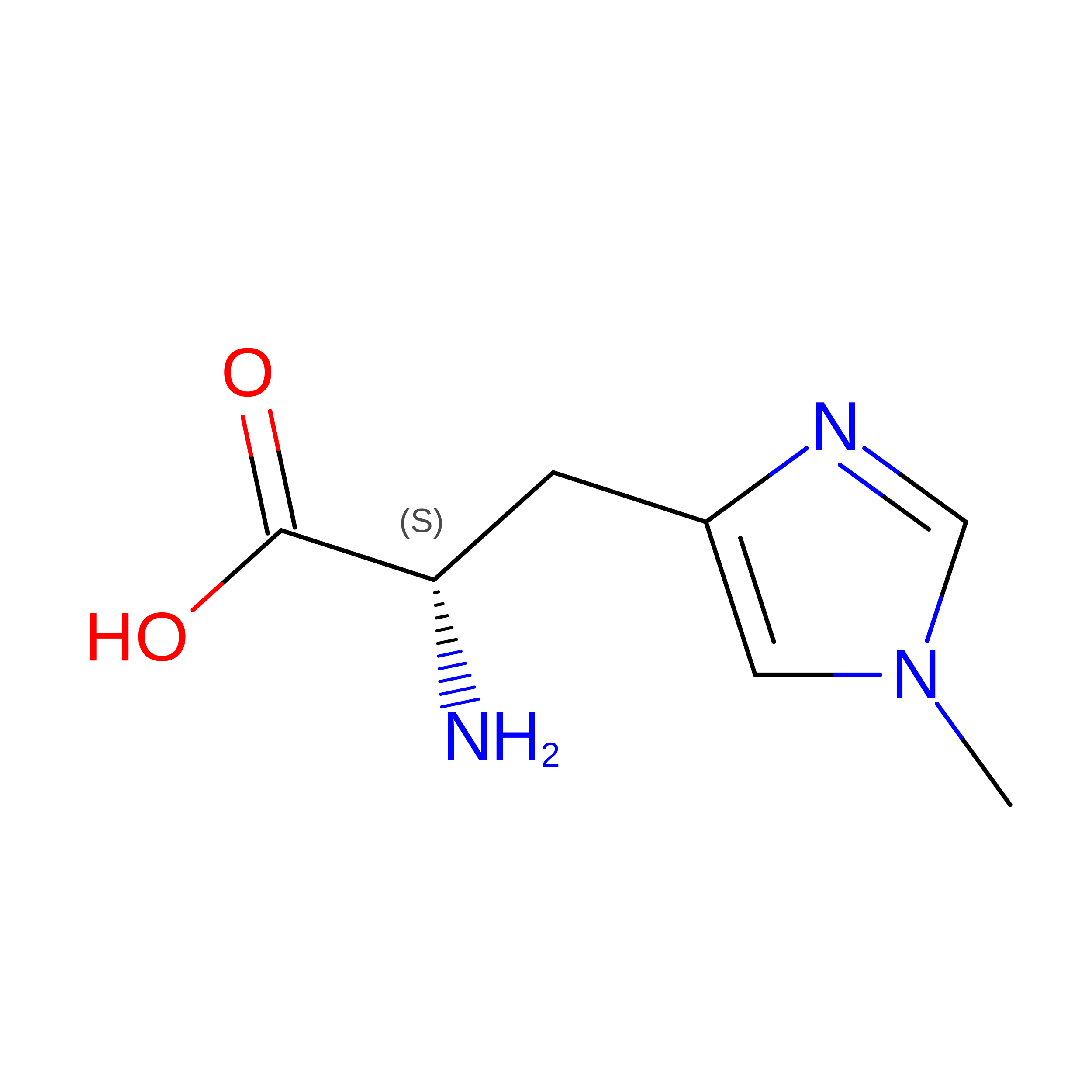
L-PEPTIDE LINKINGC7 H11 N3 O2HIS

Experimental Data & Validation

Experimental Data

  • Method: ELECTRON MICROSCOPY
  • Resolution: 3.28 Å
  • Resolution: 3.28 Å
  • Resolution: 3.28 Å
  • Resolution: 3.28 Å
  • Resolution: 3.28 Å
  • Resolution: 3.28 Å
  • Resolution: 3.28 Å
  • Resolution: 3.28 Å
  • Aggregation State: FILAMENT 
  • Reconstruction Method: SINGLE PARTICLE 
EM Software:
TaskSoftware PackageVersion
MODEL REFINEMENTPHENIX
RECONSTRUCTIONPHENIX

Structure Validation

View Full Validation Report


Entry History 

& Funding Information

Deposition Data


Funding OrganizationLocationGrant Number
National Institutes of Health/National Institute of General Medical Sciences (NIH/NIGMS)United StatesR35 GM161161
National Institutes of Health/National Institute of Arthritis and Musculoskeletal and Skin Diseases (NIH/NIAMS)United States5T32AR053461-20

Revision History  (Full details and data files)

  • Version 1.0: 2026-06-24
    Type: Initial release
  • Version 1.1: 2026-08-05
    Changes: Data collection, Database references