
 Download Mendeley
Crystal structure of macrodomain from Eastern Equine Encephalitis Virus in complex with Adenosine diphosphate ribose
Chang, C., Endre, M., Stols, L., Kim, Y., Joachimiak, A.To be published.
Experimental Data Snapshot
Starting Model: experimental
View more details
Entity ID: 1 | |||||
|---|---|---|---|---|---|
| Molecule | Chains | Sequence Length | Organism | Details | Image |
| Polyprotein P1234 | 158 | Eastern equine encephalitis virus | Mutation(s): 0 | 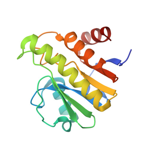 | |
UniProt | |||||
Entity Groups | |||||
| Sequence Clusters | 30% Identity50% Identity70% Identity90% Identity95% Identity100% Identity | ||||
| UniProt Group | Q66580 | ||||
Sequence AnnotationsExpand | |||||
Reference Sequence | |||||
| Ligands 3 Unique | |||||
|---|---|---|---|---|---|
| ID | Chains | Name / Formula / InChI Key | 2D Diagram | 3D Interactions | |
| AR6 Download:Ideal Coordinates CCD File | D [auth A] | [(2R,3S,4R,5R)-5-(6-AMINOPURIN-9-YL)-3,4-DIHYDROXY-OXOLAN-2-YL]METHYL[HYDROXY-[[(2R,3S,4R,5S)-3,4,5-TRIHYDROXYOXOLAN-2-YL]METHOXY]PHOSPHORYL] HYDROGEN PHOSPHATE C15 H23 N5 O14 P2 SRNWOUGRCWSEMX-ZQSHOCFMSA-N | 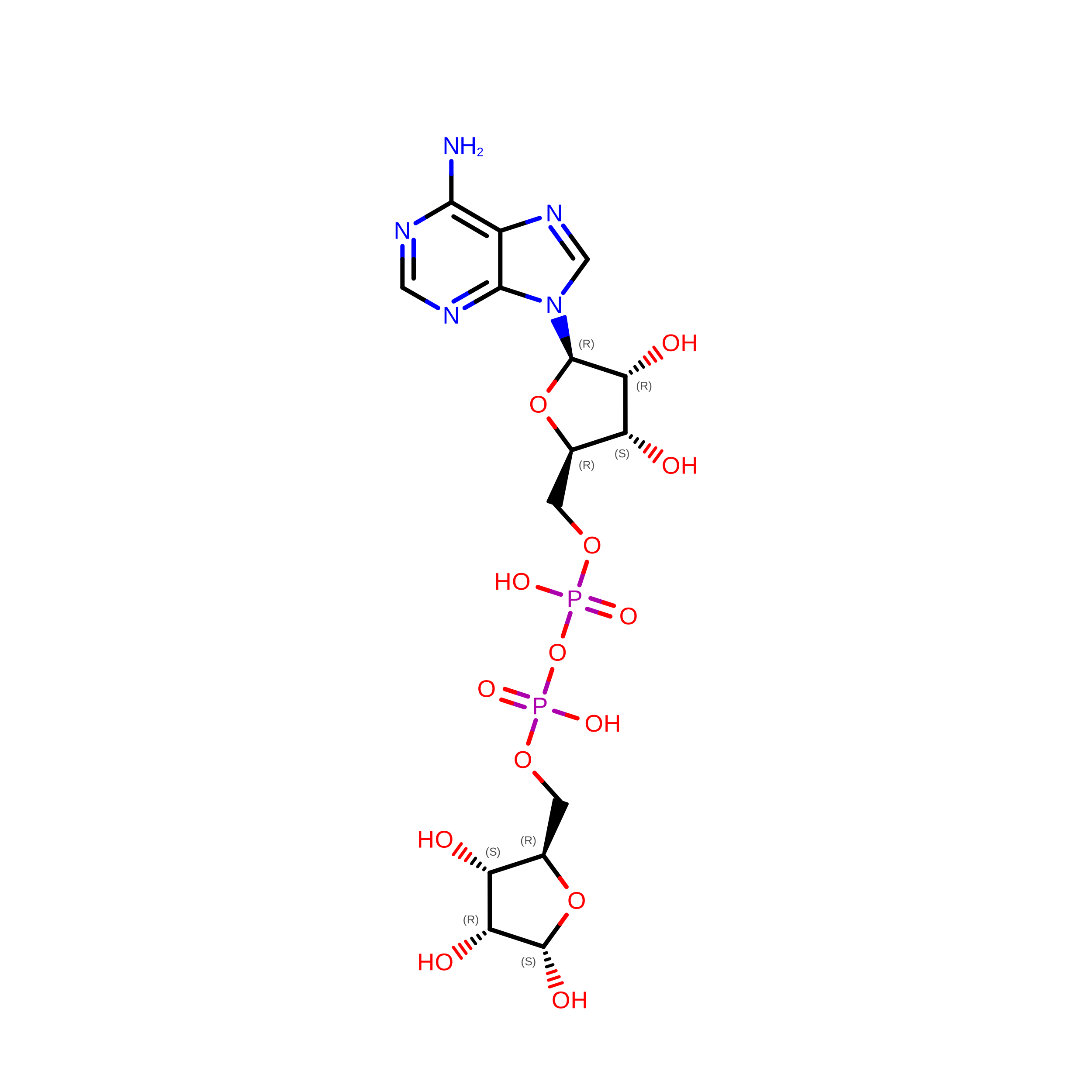 | ||
| APR (Subject of Investigation/LOI) Download:Ideal Coordinates CCD File | C [auth A] | ADENOSINE-5-DIPHOSPHORIBOSE C15 H23 N5 O14 P2 SRNWOUGRCWSEMX-KEOHHSTQSA-N | 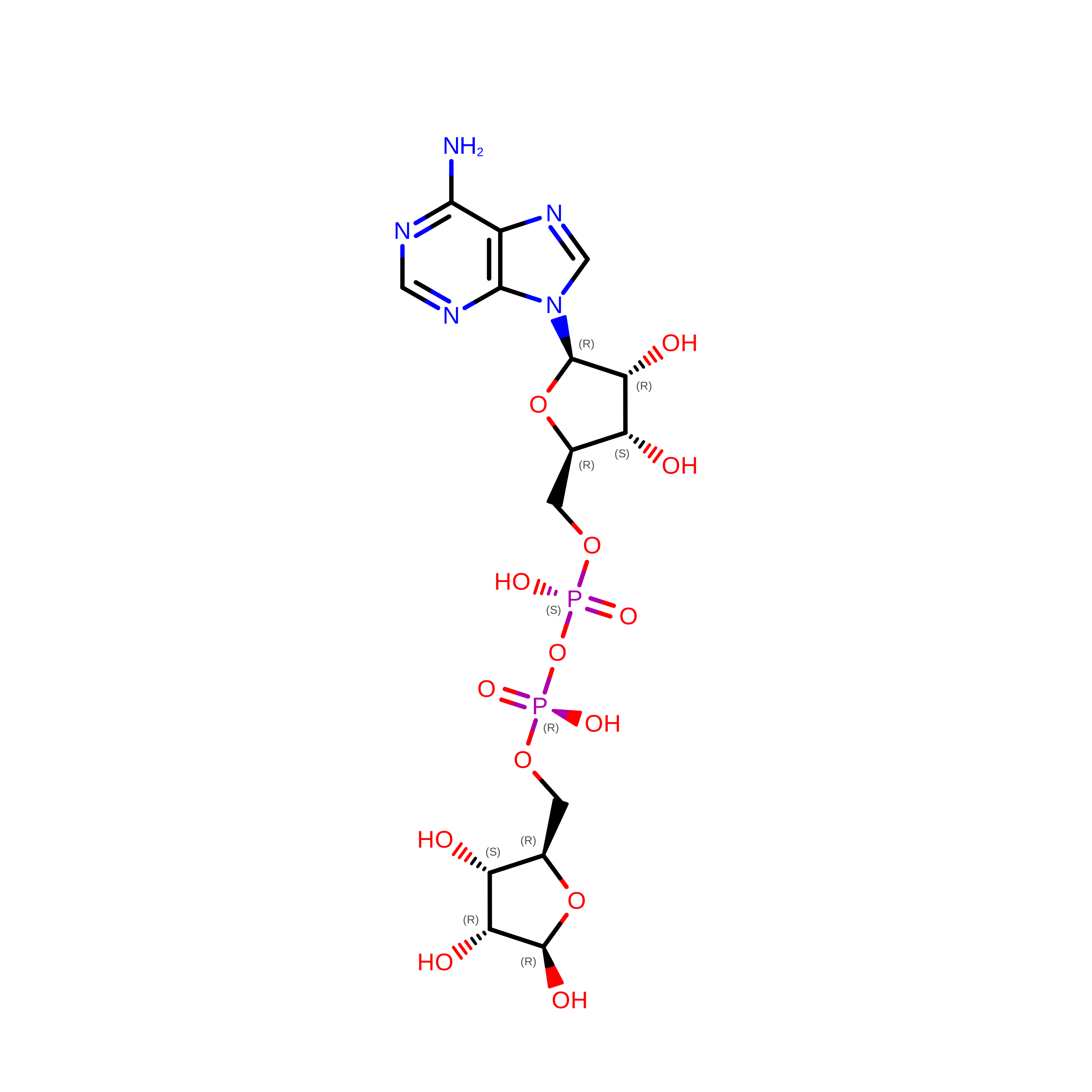 | ||
| SO4 Download:Ideal Coordinates CCD File | B [auth A] | SULFATE ION O4 S QAOWNCQODCNURD-UHFFFAOYSA-L |  | ||
| Length ( Å ) | Angle ( ˚ ) |
|---|---|
| a = 39.925 | α = 90 |
| b = 47.462 | β = 90 |
| c = 66.421 | γ = 90 |
| Software Name | Purpose |
|---|---|
| PHENIX | refinement |
| HKL-3000 | data scaling |
| PDB_EXTRACT | data extraction |
| HKL-3000 | data reduction |
| HKL-3000 | phasing |
| Funding Organization | Location | Grant Number |
|---|---|---|
| Department of Energy (DOE, United States) | United States | -- |
