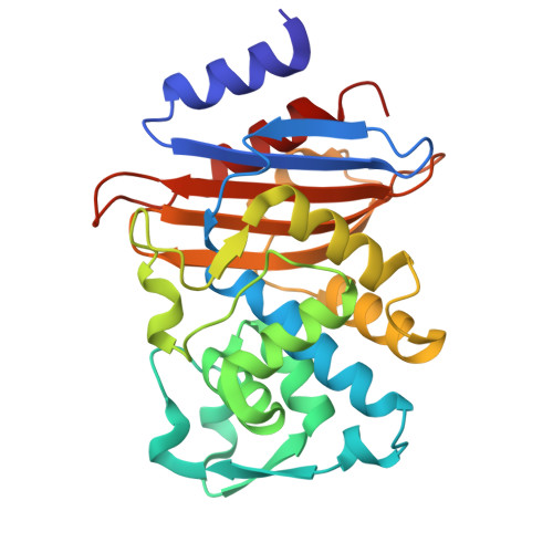
9ZQQ | pdb_00009zqq

Toho1-Avibactam X-ray structure


Experimental Data Snapshot

  • Method: X-RAY DIFFRACTION
  • Resolution: 2.00 Å
  • R-Value Free: 
    0.254 (Depositor), 0.255 (DCC) 
  • R-Value Work: 
    0.199 (Depositor), 0.199 (DCC) 
  • R-Value Observed: 
    0.202 (Depositor) 

Starting Model: experimental
View more details

wwPDB Validation 3D Report Full Report

Validation slider image for 9ZQQ

Ligand Structure Quality Assessment 


This is version 1.0 of the entry. See complete history. 

Literature

NMR crystallography reveals active-site protonation states of Toho-1 beta-lactamase in complex with avibactam.

Williams, C.G., Wang, S., Carta, V., Langan, P.S., Thome, A.F., Ramos, S.A., Holmes, J.B., Ghosh, R.K., Weiss, K.L., Beran, G.J.O., Hartman, J.D., Coates, L., Rienstra, C.M., Mueller, L.J.

(2026) Proc Natl Acad Sci U S A 123: e2616547123-e2616547123

  • DOI: https://doi.org/10.1073/pnas.2616547123
  • Primary Citation Related Structures: 
    9ZO2, 9ZOZ, 9ZQQ

  • PubMed Abstract: 

    The determination of active-site protonation states is critical for a full mechanistic understanding of enzyme catalysis and inhibition. Here, we employ NMR crystallography-the integrated combination of solid-state NMR spectroscopy, X-ray diffraction, and first-principles computational chemistry-to determine the protonation states of the active site of Toho-1 β-lactamase in complex with the non-β-lactam inhibitor avibactam. We report two X-ray crystal structures of the Toho-1:avibactam complex, along with high-field solid-state NMR measurements that enable near-complete backbone and side-chain resonance assignments. To overcome the computational scaling limits that have traditionally hindered NMR crystallography in large systems, we use an accelerated workflow in which machine-learning interatomic potentials enable efficient geometry refinement prior to density functional theory chemical shift calculations. For Toho-1, quantitative analysis of the active-site chemical shifts and chemical shift tensors using this hybrid protocol reveals that the key active-site side chains retain their canonical charge states in the presence of avibactam, with Lys73 and Lys234 protonated and positively charged, and Glu166 deprotonated and poised to function as a general base. Contrary to recent proposals suggesting that avibactam inhibits by suppressing essential proton transfers through p K a perturbations, our data point to a more direct chemical origin arising from the intrinsic resistance of the Ser70-avibactam carbamoyl linkage to hydrolysis.


  • Organizational Affiliation: 
    • Department of Chemistry, University of California-Riverside, Riverside, CA 92521.

Macromolecule Content 

  • Total Structure Weight: 28.64 kDa 
  • Atom Count: 2,217 
  • Modeled Residue Count: 261 
  • Deposited Residue Count: 261 
  • Unique protein chains: 1

Macromolecules

Find similar proteins by:|  3D Structure
Entity ID: 1
MoleculeChains  Sequence LengthOrganismDetailsImage
Beta-lactamase261Escherichia coliMutation(s): 0 
Gene Names: bla CTX-M-56
EC: 3.5.2.6
UniProt
Find proteins for A3FFV8 (Escherichia coli)
Explore A3FFV8 
Go to UniProtKB:  A3FFV8
Entity Groups
Sequence Clusters30% Identity50% Identity70% Identity90% Identity95% Identity100% Identity
UniProt GroupA3FFV8
Sequence Annotations
Expand
Reference Sequence

Small Molecules

Ligands 2 Unique
IDChains Name / Formula / InChI Key2D Diagram3D Interactions
NXL
(Subject of Investigation/LOI)

Query on NXL


Download:Ideal Coordinates CCD File
B [auth A](2S,5R)-1-formyl-5-[(sulfooxy)amino]piperidine-2-carboxamide
C7 H13 N3 O6 S
WJDGWXPPFHLLNL-RITPCOANSA-N
FYG

Query on FYG


Download:Ideal Coordinates CCD File
C [auth A](2S,5R)-7-oxo-6-(sulfooxy)-1,6-diazabicyclo[3.2.1]octane-2-carboxamide
C7 H11 N3 O6 S
NDCUAPJVLWFHHB-UHNVWZDZSA-N

Experimental Data & Validation

Experimental Data

  • Method: X-RAY DIFFRACTION
  • Resolution: 2.00 Å
  • R-Value Free:  0.254 (Depositor), 0.255 (DCC) 
  • R-Value Work:  0.199 (Depositor), 0.199 (DCC) 
  • R-Value Observed: 0.202 (Depositor) 
Space Group: P 21 21 21
Unit Cell:
Length ( Å )Angle ( ˚ )
a = 44.834α = 90
b = 73.295β = 90
c = 78.391γ = 90
Software Package:
Software NamePurpose
PHENIXrefinement
SAINTdata reduction
Aimlessdata scaling
PHASERphasing

Structure Validation

View Full Validation Report


Ligand Structure Quality Assessment 


Entry History 

& Funding Information

Deposition Data


Funding OrganizationLocationGrant Number
National Institutes of Health/National Institute of General Medical Sciences (NIH/NIGMS)United StatesR35GM145369

Revision History  (Full details and data files)

  • Version 1.0: 2026-10-28
    Type: Initial release