9Z97 | pdb_00009z97

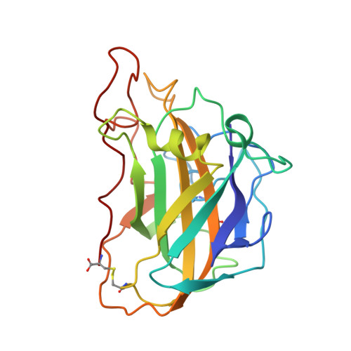
Neurospora crassa polysaccharide monooxygenase 9D dose series - pseudohelix 19 (5.91 MGy)


Experimental Data Snapshot

  • Method: X-RAY DIFFRACTION
  • Resolution: 1.10 Å
  • R-Value Free: 
    0.193 (Depositor), 0.194 (DCC) 
  • R-Value Work: 
    0.172 (Depositor), 0.172 (DCC) 
  • R-Value Observed: 
    0.172 (Depositor) 

Starting Model: experimental
View more details

wwPDB Validation 3D Report Full Report

Validation slider image for 9Z97

Ligand Structure Quality Assessment 


This is version 1.0 of the entry. See complete history

Literature

Dose-dependent structural and electron-density features in the lytic polysaccharide monooxygenase NcAA9D.

Miller, S.A.O'Dell, W.B.Meilleur, F.

(2026) Acta Crystallogr D Struct Biol 82: 900-914

  • DOI: https://doi.org/10.1107/S205979832600639X
  • Primary Citation Related Structures: 
    9Z8O, 9Z8P, 9Z8Q, 9Z8R, 9Z8S, 9Z8T, 9Z8U, 9Z8V, 9Z8W, 9Z8X, 9Z8Y, 9Z8Z, 9Z90, 9Z92, 9Z93, 9Z94, 9Z95, 9Z96, 9Z97, 9Z98, 9Z99, 9ZA5, 9ZA6, 9ZA7, 9ZA8, 9ZA9, 9ZAA, 9ZAB, 9ZAC, 9ZAD, 9ZAE, 9ZAF, 9ZAH, 9ZAI, 9ZAJ, 9ZAL, 9ZAM, 9ZAN, 9ZAQ, 9ZAR, 9ZAS

  • PubMed Abstract: 

    Structural studies of copper-containing lytic polysaccharide monooxygenases (LPMOs) by X-ray crystallography are often complicated by radiation damage. In this study, we analyze a series of 36 X-ray crystal structures of NcAA9D, a Neurospora crassa AA9-family LPMO, determined from data collected at cryogenic temperature from a single crystal to investigate the progressive effects of radiation damage at the active site of this enzyme. We report new insights into the dose-dependence of active-site geometry in LPMOs and utilize the unique pre-bound dioxygen site of NcAA9D to analyze the impact of X-ray dose on the electron density of this species. It is well established that photoreduction of the LPMO active-site copper(II) leads to expulsion of its water ligands. We further characterize this displacement and the corresponding electron-density smearing, a phenomenon that can lead to the erroneous modeling of copper-bound dioxygen species. These findings suggest that radiation-dose series collected from a single crystal provide invaluable data to support unambiguous assignment of radiation-sensitive intermediates at the active site of LPMOs and other radiation-sensitive redox enzymes.


  • Organizational Affiliation
    • Department of Molecular and Structural Biochemistry, North Carolina State University, 128 Polk Hall, Raleigh, NC 27695, USA.

Macromolecule Content 

  • Total Structure Weight: 48.51 kDa 
  • Atom Count: 5,101 
  • Modeled Residue Count: 446 
  • Deposited Residue Count: 446 
  • Unique protein chains: 1

Macromolecules

Find similar proteins by:|  3D Structure
Entity ID: 1
MoleculeChains  Sequence LengthOrganismDetailsImage
Lytic polysaccharide monooxygenase NCU01050
A, B
223Neurospora crassaMutation(s): 0 
Gene Names: gh61-4NCU01050
EC: 1.14.99.56
UniProt
Find proteins for Q1K8B6 (Neurospora crassa (strain ATCC 24698 / 74-OR23-1A / CBS 708.71 / DSM 1257 / FGSC 987))
Explore Q1K8B6 
Go to UniProtKB:  Q1K8B6
Entity Groups
Sequence Clusters30% Identity50% Identity70% Identity90% Identity95% Identity100% Identity
UniProt GroupQ1K8B6
Glycosylation
Glycosylation Sites: 1
Sequence Annotations
Expand
Reference Sequence

Oligosaccharides

Help  
Entity ID: 2
MoleculeChains Length2D Diagram GlycosylationD Interactions
beta-D-mannopyranose-(1-4)-2-acetamido-2-deoxy-beta-D-glucopyranose-(1-4)-2-acetamido-2-deoxy-beta-D-glucopyranose
C
3N-Glycosylation
Entity ID: 3
MoleculeChains Length2D Diagram GlycosylationD Interactions
2-acetamido-2-deoxy-beta-D-glucopyranose-(1-4)-2-acetamido-2-deoxy-beta-D-glucopyranose
D
2N-Glycosylation

Small Molecules

Ligands 5 Unique
IDChains Name / Formula / InChI Key2D Diagram3D Interactions
PEG

Query on PEG


Download:Ideal Coordinates CCD File
I [auth A]DI(HYDROXYETHYL)ETHER
C4 H10 O3
MTHSVFCYNBDYFN-UHFFFAOYSA-N
CU
(Subject of Investigation/LOI)

Query on CU


Download:Ideal Coordinates CCD File
K [auth A],
S [auth B]
COPPER (II) ION
Cu
JPVYNHNXODAKFH-UHFFFAOYSA-N
EDO

Query on EDO


Download:Ideal Coordinates CCD File
E [auth A]
F [auth A]
G [auth A]
H [auth A]
J [auth A]
E [auth A],
F [auth A],
G [auth A],
H [auth A],
J [auth A],
O [auth B],
P [auth B],
Q [auth B],
R [auth B]
1,2-ETHANEDIOL
C2 H6 O2
LYCAIKOWRPUZTN-UHFFFAOYSA-N
CO2
(Subject of Investigation/LOI)

Query on CO2


Download:Ideal Coordinates CCD File
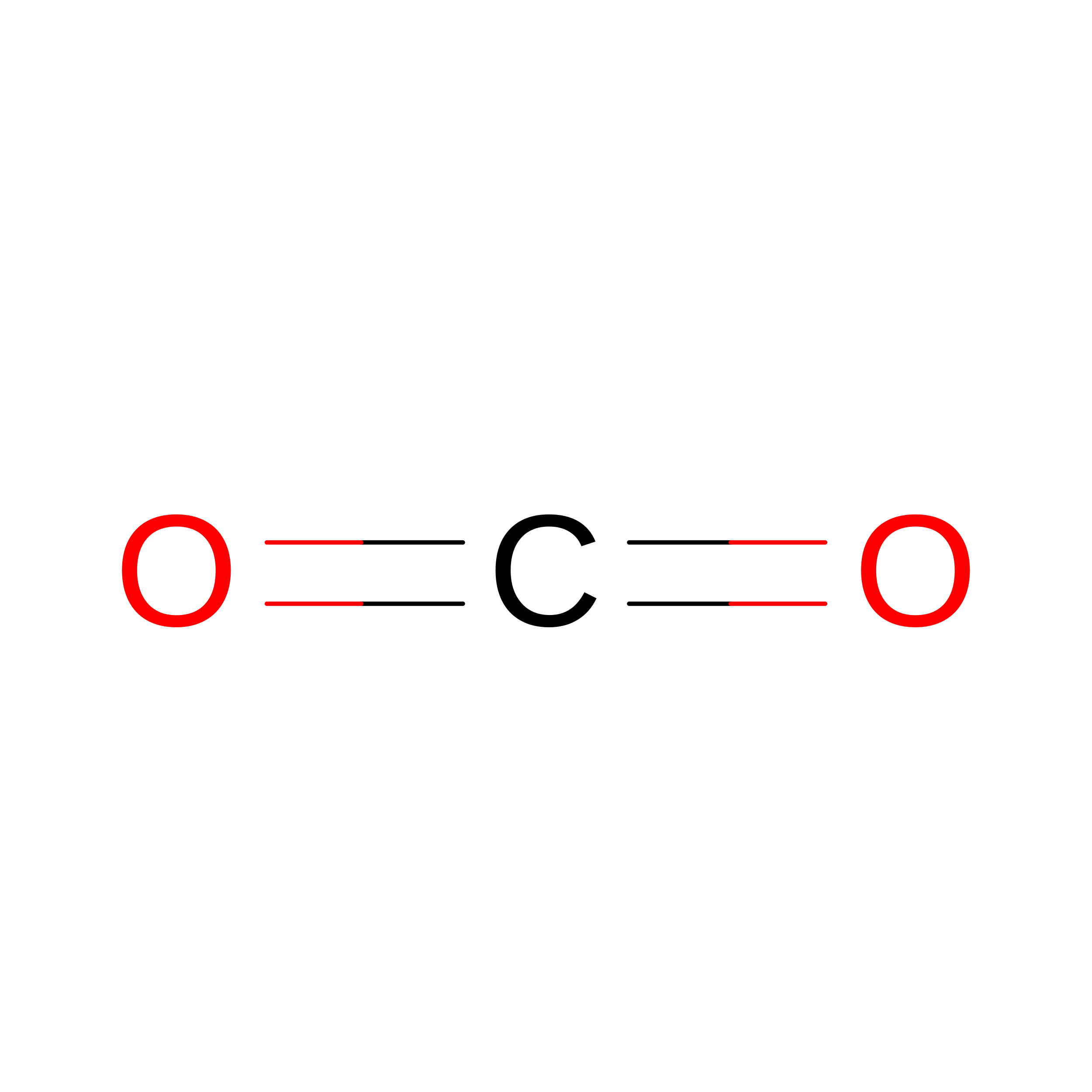
N [auth A]CARBON DIOXIDE
C O2
CURLTUGMZLYLDI-UHFFFAOYSA-N
OXY
(Subject of Investigation/LOI)

Query on OXY


Download:Ideal Coordinates CCD File
L [auth A],
M [auth A]
OXYGEN MOLECULE
O2
MYMOFIZGZYHOMD-UHFFFAOYSA-N

Experimental Data & Validation

Experimental Data

  • Method: X-RAY DIFFRACTION
  • Resolution: 1.10 Å
  • R-Value Free:  0.193 (Depositor), 0.194 (DCC) 
  • R-Value Work:  0.172 (Depositor), 0.172 (DCC) 
  • R-Value Observed: 0.172 (Depositor) 
Space Group: P 1 21 1
Unit Cell:
Length ( Å )Angle ( ˚ )
a = 67.522α = 90
b = 42.234β = 98.988
c = 69.58γ = 90
Software Package:
Software NamePurpose
DIALSdata reduction
DIALSdata scaling
PHENIXphasing
Cootmodel building
PHENIXrefinement

Structure Validation

View Full Validation Report


Ligand Structure Quality Assessment 


Entry History 

& Funding Information

Deposition Data


Funding OrganizationLocationGrant Number
Department of Energy (DOE, United States)United States--

Revision History  (Full details and data files)

  • Version 1.0: 2026-08-12
    Type: Initial release