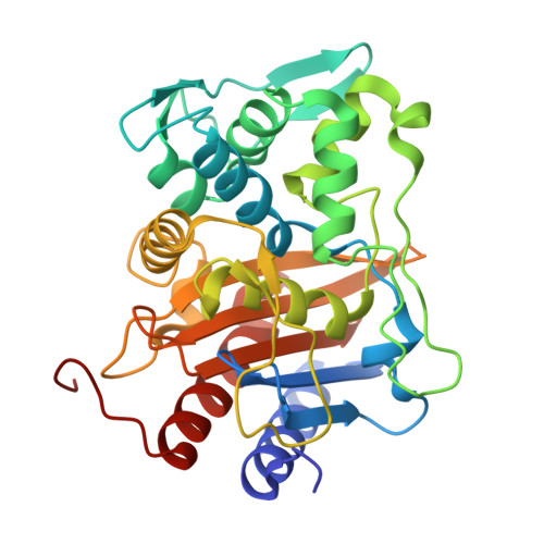
9Z0X | pdb_00009z0x

Crystal structure of Neisseria gonorrhoeae penicillin-binding protein 2 from strain FA19 containing seven resistance mutations

  • Classification: HYDROLASE
  • Organism(s): Neisseria gonorrhoeae FA19
  • Expression System: Escherichia coli BL21(DE3)
  • Mutation(s): Yes 

  • Deposited: 2025-11-03 Released: 2026-03-04 
  • Deposition Author(s): Singh, A., Bala, S., Davies, C.
  • Funding Organization(s): National Institutes of Health/National Institute Of Allergy and Infectious Diseases (NIH/NIAID), National Institutes of Health/National Institute of General Medical Sciences (NIH/NIGMS)

Experimental Data Snapshot

  • Method: X-RAY DIFFRACTION
  • Resolution: 1.90 Å
  • R-Value Free: 
    0.239 (Depositor), 0.244 (DCC) 
  • R-Value Work: 
    0.200 (Depositor), 0.204 (DCC) 
  • R-Value Observed: 
    0.202 (Depositor) 

Starting Model: experimental
View more details

wwPDB Validation 3D Report Full Report

Validation slider image for 9Z0X

This is version 1.1 of the entry. See complete history

Literature

Antibiotic-resistance mutations in penicillin-binding protein 2 from the ceftriaxone-resistant Neisseria gonorrhoeae strain H041 strike a delicate balance between increasing resistance and maintaining transpeptidase activity.

Bivins, M.M.Tomberg, J.Bagshaw, M.Singh, A.Bala, S.Davies, C.Nicholas, R.A.

(2026) PLoS Pathog 22: e1013721-e1013721

  • DOI: https://doi.org/10.1371/journal.ppat.1013721
  • Primary Citation Related Structures: 
    9Z0W, 9Z0X, 9Z0Y

  • PubMed Abstract: 

    The mosaic penA allele (penA41) from H041, the most ceftriaxone-resistant Neisseria gonorrhoeae strain identified to date, encodes a variant of the essential Penicillin-Binding Protein 2 (PBP2) with 60 amino acid mutations compared to PBP2 from the antimicrobial-susceptible strain, FA19. Based on previous work from our lab and others, we identified a minimal set of 10 mutations that, when introduced into the β-lactam antibiotic-susceptible penA allele from FA19 (penA19), confers two-thirds of the ceftriaxone and cefixime resistance compared to the penA41 allele. Three mutations (A311V, I312M, and V316P) are in the α2 helix of PBP2 containing the catalytic serine (Ser310), two (F504L and N512Y) are in the β3-β4 loop that is important in binding and acylation, and one (G545S) interacts with conserved amino acids in the active site. The seventh mutation, T483S, confers substantial resistance to ceftriaxone within the minimal mutant set but requires the presence of three epistatic mutations located on the other side of the protein that do not alter resistance on their own yet are necessary to retain essential function. These epistatic mutations change the backbone dihedral angles at position-447, which may increase flexibility of the enzyme and help maintain enzymatic function. Our results highlight the complex balance necessary for evolving cephalosporin resistance in PBP2 while also retaining sufficient transpeptidase function to support growth.


  • Organizational Affiliation
    • Department of Pharmacology, University of North Carolina at Chapel Hill, Chapel Hill, North Carolina, United States of America.

Macromolecule Content 

  • Total Structure Weight: 70.85 kDa 
  • Atom Count: 5,015 
  • Modeled Residue Count: 642 
  • Deposited Residue Count: 658 
  • Unique protein chains: 1

Macromolecules

Find similar proteins by:|  3D Structure
Entity ID: 1
MoleculeChains  Sequence LengthOrganismDetailsImage
Penicillin-binding protein 2
A, B
329Neisseria gonorrhoeae FA19Mutation(s): 7 
Gene Names: penAWHOF_00731WHOF_01799C
EC: 3.4.16.4
UniProt
Find proteins for A0AB74EE38 (Neisseria gonorrhoeae)
Explore A0AB74EE38 
Go to UniProtKB:  A0AB74EE38
Entity Groups
Sequence Clusters30% Identity50% Identity70% Identity90% Identity95% Identity100% Identity
UniProt GroupA0AB74EE38
Sequence Annotations
Expand
Reference Sequence

Small Molecules

Ligands 1 Unique
IDChains Name / Formula / InChI Key2D Diagram3D Interactions
PO4

Query on PO4


Download:Ideal Coordinates CCD File
C [auth B]PHOSPHATE ION
O4 P
NBIIXXVUZAFLBC-UHFFFAOYSA-K

Experimental Data & Validation

Experimental Data

  • Method: X-RAY DIFFRACTION
  • Resolution: 1.90 Å
  • R-Value Free:  0.239 (Depositor), 0.244 (DCC) 
  • R-Value Work:  0.200 (Depositor), 0.204 (DCC) 
  • R-Value Observed: 0.202 (Depositor) 
Space Group: P 1 21 1
Unit Cell:
Length ( Å )Angle ( ˚ )
a = 44.556α = 90
b = 76.839β = 92.37
c = 87.478γ = 90
Software Package:
Software NamePurpose
REFMACrefinement
HKL-2000data scaling
HKL-2000data reduction
PDB_EXTRACTdata extraction
REFMACphasing

Structure Validation

View Full Validation Report


Entry History 

& Funding Information

Deposition Data


Funding OrganizationLocationGrant Number
National Institutes of Health/National Institute Of Allergy and Infectious Diseases (NIH/NIAID)United StatesAI164794-01
National Institutes of Health/National Institute of General Medical Sciences (NIH/NIGMS)United StatesGM066861

Revision History  (Full details and data files)

  • Version 1.0: 2026-03-04
    Type: Initial release
  • Version 1.1: 2026-09-16
    Changes: Database references