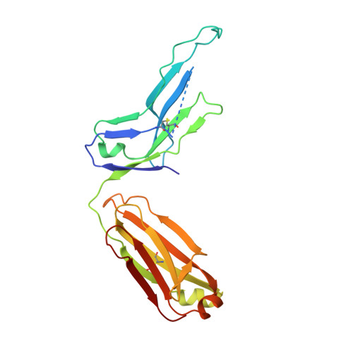
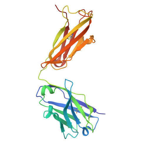
9Z0T | pdb_00009z0t

Fab structure of anti-Tau mAb G10

  • Classification: IMMUNE SYSTEM
  • Organism(s): Mus musculus
  • Expression System: Homo sapiens
  • Mutation(s): No 

  • Deposited: 2025-11-03 Released: 2026-10-14 
  • Deposition Author(s): Pan, R., Kong, X.P.
  • Funding Organization(s): National Institutes of Health/National Institute of Neurological Disorders and Stroke (NIH/NINDS), National Institutes of Health/National Institute on Aging (NIH/NIA)

Experimental Data Snapshot

  • Method: X-RAY DIFFRACTION
  • Resolution: 3.02 Å
  • R-Value Free: 
    0.252 (Depositor), 0.250 (DCC) 
  • R-Value Work: 
    0.196 (Depositor), 0.199 (DCC) 
  • R-Value Observed: 
    0.199 (Depositor) 

Starting Model: experimental
View more details

wwPDB Validation 3D Report Full Report

Validation slider image for 9Z0T

This is version 1.1 of the entry. See complete history. 

Literature

Tau pSer396 and pSer404 Define Distinct Epitope Regions Linked to Different Antibody Functions.

Pan, R., Congdon, E.E., Chukwu, J.E., Luo, C.C., Sigurdsson, E.M., Kong, X.P.

(2026) bioRxiv 

  • DOI: https://doi.org/10.64898/2026.04.16.716390
  • Primary Citation Related Structures: 
    9Z0S, 9Z0T, 9Z0U, 9Z0V

  • PubMed Abstract: 

    Hyperphosphorylated tau is a central pathological feature of Alzheimer's disease and related tauopathies, and antibodies targeting the pSer396/pSer404 epitope region represent a promising therapeutic strategy. However, direct comparisons of pSer396- and pSer404-selective antibodies and the impact of humanization on their functional properties remain limited. We generated two new monoclonal antibodies (mAbs), 9E (pSer404-specific) and G10 (pSer396-specific), and evaluated them alongside 4E6 (pSer404) and PHF-1 (pSer396) in murine and partially humanized chimeric formats. Antibodies were assessed in mixed cortical cultures using extracellular (PHF + Ab) and intracellular (PHF → Ab) paradigms. Efficacy in preventing tau-induced toxicity and seeding differed substantially among antibodies and was variably altered by chimerization, despite identical variable regions. Antibodies targeting pSer404 were more effective than those targeting pSer396, and antibodies that preferentially bound soluble pathological tau species in competition ELISA were consistently more efficacious, whereas neuronal uptake was comparable across variants. To define structural determinants of phospho-epitope recognition, we determined the crystal structures of the Fab regions of 9E, G10, and PHF-1, and additionally solved the co-crystal structure of Fab PHF-1 in complex with a pSer396 tau peptide at 2.55 Å resolution. The PHF-1 complex reveals a heavy-chain-dominant binding mode in which pSer396 is anchored within an electropositive pocket and Tyr394 adopts a flipped conformation that stabilizes a β-strand-like motif, consistent with a phosphorylation-dependent conformational switch. These findings demonstrate that epitope selectivity, aggregate preference, structural binding mode, and Fc context collectively govern antibody efficacy, and that humanization can substantially alter therapeutic properties.


  • Organizational Affiliation: 
    • Department of Biochemistry and Molecular Pharmacology, New York University Grossman School of Medicine, New York NY 10016, USA.

Macromolecule Content 

  • Total Structure Weight: 93.09 kDa 
  • Atom Count: 6,414 
  • Modeled Residue Count: 843 
  • Deposited Residue Count: 860 
  • Unique protein chains: 2

Macromolecules

Find similar proteins by:|  3D Structure
Entity ID: 1
MoleculeChains  Sequence LengthOrganismDetailsImage
Light chain of Fab fragment of anti-Tau mAb G10A [auth L],
C [auth M]
211Mus musculusMutation(s): 0 
Entity Groups
Sequence Clusters30% Identity50% Identity70% Identity90% Identity95% Identity100% Identity
Sequence Annotations
Expand
Reference Sequence
Find similar proteins by:|  3D Structure
Entity ID: 2
MoleculeChains  Sequence LengthOrganismDetailsImage
Heavy chain of Fab fragment of anti-tau mAb G10B [auth H],
D [auth I]
219Mus musculusMutation(s): 0 
Entity Groups
Sequence Clusters30% Identity50% Identity70% Identity90% Identity95% Identity100% Identity
Sequence Annotations
Expand
Reference Sequence

Experimental Data & Validation

Experimental Data

  • Method: X-RAY DIFFRACTION
  • Resolution: 3.02 Å
  • R-Value Free:  0.252 (Depositor), 0.250 (DCC) 
  • R-Value Work:  0.196 (Depositor), 0.199 (DCC) 
  • R-Value Observed: 0.199 (Depositor) 
Space Group: P 21 2 21
Unit Cell:
Length ( Å )Angle ( ˚ )
a = 85.642α = 90
b = 105.289β = 90
c = 132.108γ = 90
Software Package:
Software NamePurpose
PHENIXrefinement
XDSdata reduction
XDSdata scaling
PHENIXphasing

Structure Validation

View Full Validation Report


Entry History 

& Funding Information

Deposition Data


Funding OrganizationLocationGrant Number
National Institutes of Health/National Institute of Neurological Disorders and Stroke (NIH/NINDS)United StatesR01 NS120488
National Institutes of Health/National Institute of Neurological Disorders and Stroke (NIH/NINDS)United StatesR01 NS077239
National Institutes of Health/National Institute on Aging (NIH/NIA)United StatesR01 AG032611
National Institutes of Health/National Institute on Aging (NIH/NIA)United StatesR21 AG059391

Revision History  (Full details and data files)

  • Version 1.0: 2026-10-14
    Type: Initial release
  • Version 1.1: 2026-10-21
    Changes: Database references