9YR7 | pdb_00009yr7

Cryo-EM structure of human beta-cardiac myosin bound to mavacamten in the interacting-heads motif and S2-FH undocked state


Experimental Data Snapshot

  • Method: ELECTRON MICROSCOPY
  • Resolution: 3.00 Å
  • Aggregation State: PARTICLE 
  • Reconstruction Method: SINGLE PARTICLE 

Starting Models: experimental
View more details

wwPDB Validation 3D Report Full Report

Validation slider image for 9YR7

This is version 1.1 of the entry. See complete history

Literature

Cryo-EM reveals how cardiomyopathy therapeutic drugs modulate the myosin motors of the heart.

Somavarapu, A.K.Ge, J.Yengo, C.M.Craig, R.Padron, R.

(2026) Sci Adv 12: eaed6472-eaed6472

  • DOI: https://doi.org/10.1126/sciadv.aed6472
  • Primary Citation Related Structures: 
    9YOP, 9YP4, 9YP9, 9YR7, 9YRG, 9YRH

  • PubMed Abstract: 

    Genetic mutations in myosin, the motor protein that powers the heartbeat, are linked to inherited hypertrophic and dilated cardiomyopathies. Mavacamten and omecamtiv mecarbil are therapeutic, myosin-targeted drugs designed to treat these myopathies, but their mechanism of action has remained unclear. Using single-particle cryo-EM, we determined near-atomic resolution structures of wild-type, mavacamten-bound, and omecamtiv mecarbil-bound myosin molecules. Across all conditions, we observe two distinct, alternate conformations of myosin, not previously reported. We show how mavacamten stabilizes one conformation by reinforcing key electrostatic interfaces in the molecule, whereas omecamtiv mecarbil weakens these interfaces, favoring the second structure. This remodeling elucidates previously unclear allosteric mechanisms through which these drugs either inhibit or enhance myosin activity, countering the deleterious impacts of disease. These findings reveal how drugs modulate myosin structure to control cardiac contractility.


  • Organizational Affiliation
    • Division of Cell Biology and Imaging, Department of Radiology, University of Massachusetts Chan Medical School, Worcester, MA, USA.

Macromolecule Content 

  • Total Structure Weight: 382.1 kDa 
  • Atom Count: 19,808 
  • Modeled Residue Count: 2,454 
  • Deposited Residue Count: 3,344 
  • Unique protein chains: 3

Macromolecules

Find similar proteins by:|  3D Structure
Entity ID: 1
MoleculeChains  Sequence LengthOrganismDetailsImage
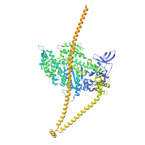
Myosin-7,General control transcription factor GCN4,Enhanced Green fluorescent protein
A, B
1,315Homo sapiensSaccharomyces cerevisiaeAequorea victoria
This entity is chimeric
Mutation(s): 4 
Gene Names: MYH7MYHCBGCN4AAS101AAS3ARG9YEL009CGFP
UniProt & NIH Common Fund Data Resources
Find proteins for P03069 (Saccharomyces cerevisiae (strain ATCC 204508 / S288c))
Explore P03069 
Go to UniProtKB:  P03069
Find proteins for P12883 (Homo sapiens)
Explore P12883 
Go to UniProtKB:  P12883
PHAROS:  P12883
GTEx:  ENSG00000092054 
Find proteins for P42212 (Aequorea victoria)
Explore P42212 
Go to UniProtKB:  P42212
Entity Groups
Sequence Clusters30% Identity50% Identity70% Identity90% Identity95% Identity100% Identity
UniProt GroupsP12883P42212P03069
Sequence Annotations
Expand
Reference Sequence
Find similar proteins by:|  3D Structure
Entity ID: 2
MoleculeChains  Sequence LengthOrganismDetailsImage
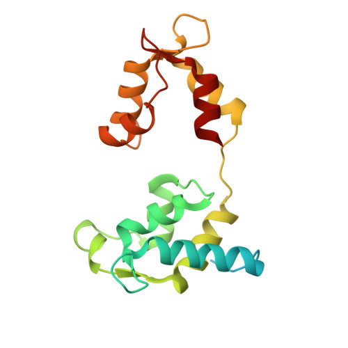
Myosin light chain 1/3, skeletal muscle isoform
C, D
188Mus musculusMutation(s): 0 
UniProt
Find proteins for P05977 (Mus musculus)
Explore P05977 
Go to UniProtKB:  P05977
Entity Groups
Sequence Clusters30% Identity50% Identity70% Identity90% Identity95% Identity100% Identity
UniProt GroupP05977
Sequence Annotations
Expand
Reference Sequence
Find similar proteins by:|  3D Structure
Entity ID: 3
MoleculeChains  Sequence LengthOrganismDetailsImage
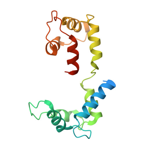
Myosin regulatory light chain 11
E, F
169Mus musculusMutation(s): 0 
UniProt
Find proteins for P97457 (Mus musculus)
Explore P97457 
Go to UniProtKB:  P97457
Entity Groups
Sequence Clusters30% Identity50% Identity70% Identity90% Identity95% Identity100% Identity
UniProt GroupP97457
Sequence Annotations
Expand
Reference Sequence

Small Molecules

Ligands 3 Unique
IDChains Name / Formula / InChI Key2D Diagram3D Interactions
ADP
(Subject of Investigation/LOI)

Query on ADP


Download:Ideal Coordinates CCD File
H [auth A],
K [auth B]
ADENOSINE-5'-DIPHOSPHATE
C10 H15 N5 O10 P2
XTWYTFMLZFPYCI-KQYNXXCUSA-N
XB2
(Subject of Investigation/LOI)

Query on XB2


Download:Ideal Coordinates CCD File
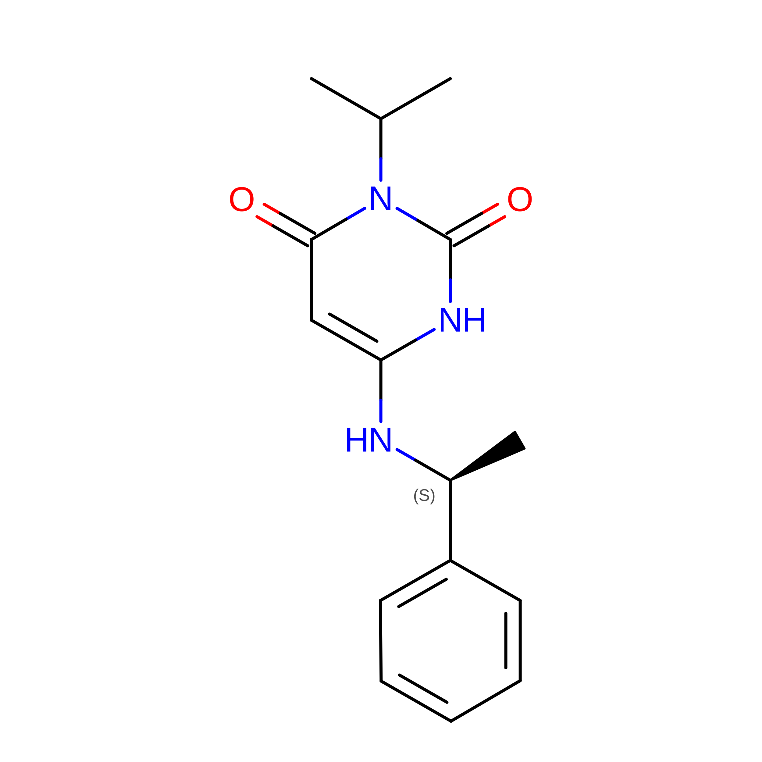
G [auth A],
J [auth B]
Mavacamten
C15 H19 N3 O2
RLCLASQCAPXVLM-NSHDSACASA-N
PO4
(Subject of Investigation/LOI)

Query on PO4


Download:Ideal Coordinates CCD File
I [auth A],
L [auth B]
PHOSPHATE ION
O4 P
NBIIXXVUZAFLBC-UHFFFAOYSA-K

Experimental Data & Validation

Experimental Data

  • Method: ELECTRON MICROSCOPY
  • Resolution: 3.00 Å
  • Aggregation State: PARTICLE 
  • Reconstruction Method: SINGLE PARTICLE 
EM Software:
TaskSoftware PackageVersion
MODEL REFINEMENTPHENIX1.20.1_4487:
RECONSTRUCTIONcryoSPARC

Structure Validation

View Full Validation Report


Entry History 

& Funding Information

Deposition Data


Funding OrganizationLocationGrant Number
National Institutes of Health/National Heart, Lung, and Blood Institute (NIH/NHLBI)United StatesHL164560
National Institutes of Health/National Institute of Arthritis and Musculoskeletal and Skin Diseases (NIH/NIAMS)United StatesAR081941

Revision History  (Full details and data files)

  • Version 1.0: 2026-04-08
    Type: Initial release
  • Version 1.1: 2026-05-20
    Changes: Data collection, Database references