9YA5 | pdb_00009ya5

KIF1A R350G bound to microtubules in two-heads-bound state with AMP-PNP


Experimental Data Snapshot

  • Method: ELECTRON MICROSCOPY
  • Resolution: 2.95 Å
  • Aggregation State: FILAMENT 
  • Reconstruction Method: HELICAL 

Starting Model: experimental
View more details

wwPDB Validation 3D Report Full Report

Validation slider image for 9YA5

This is version 1.0 of the entry. See complete history

Literature

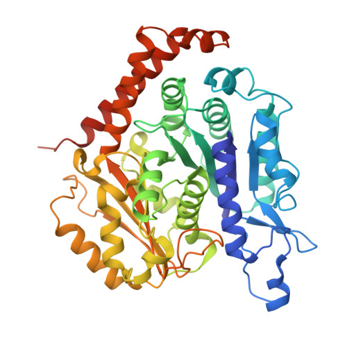
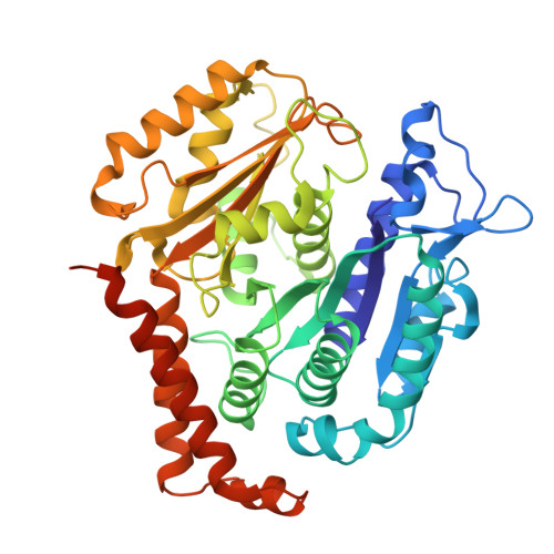
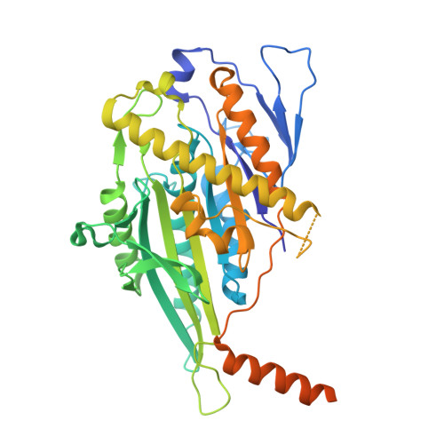
Pathogenic KIF1A R350 mutations disrupt a conserved and conformation-dependent kinesin-tubulin salt bridge.

Shatarupa, A.Rao, L.Asenjo, A.B.Gennerich, A.Sosa, H.

(2026) Nat Commun 

  • DOI: https://doi.org/10.1038/s41467-026-71026-6
  • Primary Citation Related Structures: 
    9YA5, 9YA7, 9YAB, 9YAI

  • PubMed Abstract: 

    Pathogenic variants in the motor domain of the kinesin-3 motor protein KIF1A cause a range of neurodevelopmental and neurodegenerative conditions collectively termed KIF1A-associated neurological disorder (KAND). Among these, mutations at residue R350 are linked to hereditary spastic paraplegia and altered motor function. Yet, the structural basis for their pathogenicity remains unclear. Here, we present high-resolution cryo-electron microscopy (cryo-EM) structures of KIF1A R350G and R350W bound to microtubules in both the apo and AMP-PNP-bound states. We identify a salt bridge between KIF1A residue R350 and α-tubulin E415 that forms only in the open conformation of the motor domain and is disrupted in both mutants. The loss of this electrostatic interaction correlates with increased velocity, reduced processivity, and decreased microtubule affinity in the open, apo conformation, as demonstrated by single-molecule assays. Our results reveal an electrostatic interaction at the motor-microtubule interface that regulates KIF1A's motility behavior.


  • Organizational Affiliation
    • Department of Biochemistry and Gruss-Lipper Biophotonics Center, Albert Einstein College of Medicine, Bronx, NY, USA.

Macromolecule Content 

  • Total Structure Weight: 303.51 kDa 
  • Atom Count: 19,724 
  • Modeled Residue Count: 2,475 
  • Deposited Residue Count: 2,668 
  • Unique protein chains: 3

Macromolecules

Find similar proteins by:|  3D Structure
Entity ID: 1
MoleculeChains  Sequence LengthOrganismDetailsImage
Tubulin alpha-1B chainA,
C [auth E]
451Sus scrofaMutation(s): 0 
EC: 3.6.5
UniProt
Find proteins for Q2XVP4 (Sus scrofa)
Explore Q2XVP4 
Go to UniProtKB:  Q2XVP4
Entity Groups
Sequence Clusters30% Identity50% Identity70% Identity90% Identity95% Identity100% Identity
UniProt GroupQ2XVP4
Sequence Annotations
Expand
Reference Sequence
Find similar proteins by:|  3D Structure
Entity ID: 2
MoleculeChains  Sequence LengthOrganismDetailsImage
Tubulin beta-2B chainB,
D [auth I]
445Sus scrofaMutation(s): 0 
UniProt
Find proteins for A0A287AGU7 (Sus scrofa)
Explore A0A287AGU7 
Go to UniProtKB:  A0A287AGU7
Entity Groups
Sequence Clusters30% Identity50% Identity70% Identity90% Identity95% Identity100% Identity
UniProt GroupA0A287AGU7
Sequence Annotations
Expand
Reference Sequence
Find similar proteins by:|  3D Structure
Entity ID: 3
MoleculeChains  Sequence LengthOrganismDetailsImage
Kinesin-like protein KIF1AE [auth K],
F [auth N]
438Homo sapiensMutation(s): 1 
Gene Names: KIF1AATSVC2orf20
EC: 5.6.1.3
UniProt & NIH Common Fund Data Resources
Find proteins for Q12756 (Homo sapiens)
Explore Q12756 
Go to UniProtKB:  Q12756
PHAROS:  Q12756
GTEx:  ENSG00000130294 
Entity Groups
Sequence Clusters30% Identity50% Identity70% Identity90% Identity95% Identity100% Identity
UniProt GroupQ12756
Sequence Annotations
Expand
Reference Sequence

Small Molecules

Ligands 5 Unique
IDChains Name / Formula / InChI Key2D Diagram3D Interactions
TA1
(Subject of Investigation/LOI)

Query on TA1


Download:Ideal Coordinates CCD File
J [auth B],
N [auth I]
TAXOL
C47 H51 N O14
RCINICONZNJXQF-MZXODVADSA-N
GTP

Query on GTP


Download:Ideal Coordinates CCD File
G [auth A],
K [auth E]
GUANOSINE-5'-TRIPHOSPHATE
C10 H16 N5 O14 P3
XKMLYUALXHKNFT-UUOKFMHZSA-N
ANP

Query on ANP


Download:Ideal Coordinates CCD File
P [auth K],
Q [auth N]
PHOSPHOAMINOPHOSPHONIC ACID-ADENYLATE ESTER
C10 H17 N6 O12 P3
PVKSNHVPLWYQGJ-KQYNXXCUSA-N
GDP

Query on GDP


Download:Ideal Coordinates CCD File
I [auth B],
M [auth I]
GUANOSINE-5'-DIPHOSPHATE
C10 H15 N5 O11 P2
QGWNDRXFNXRZMB-UUOKFMHZSA-N
MG

Query on MG


Download:Ideal Coordinates CCD File
H [auth A],
L [auth E],
O [auth K]
MAGNESIUM ION
Mg
JLVVSXFLKOJNIY-UHFFFAOYSA-N

Experimental Data & Validation

Experimental Data

  • Method: ELECTRON MICROSCOPY
  • Resolution: 2.95 Å
  • Aggregation State: FILAMENT 
  • Reconstruction Method: HELICAL 
EM Software:
TaskSoftware PackageVersion
MODEL REFINEMENTPHENIX
RECONSTRUCTIONcryoSPARC4.6.0

Structure Validation

View Full Validation Report


Entry History 

& Funding Information

Deposition Data


Funding OrganizationLocationGrant Number
National Institutes of Health/National Institute of General Medical Sciences (NIH/NIGMS)United StatesR01GM147332
National Institutes of Health/National Institute of General Medical Sciences (NIH/NIGMS)United StatesRO1GM113164
National Institutes of Health/National Institute of General Medical Sciences (NIH/NIGMS)United StatesGM103310

Revision History  (Full details and data files)

  • Version 1.0: 2026-04-22
    Type: Initial release