Download Mendeley
Antigen-Detected NMR for Minimal Epitope Engineering and Structure-Guided Selection of a Na V 1.7-Selective Nanobody.
Liu, J., Chen, W., Cristofori-Armstrong, B., Crawford, T., Chen, K.E., Xie, P., Chan, R.W.B., Zhu, Y., Golder, M., Pereira Schmidt, A., Naughton, J.D., Condon, N.D., Andersson, A., Dehkhoda, F., McMahon, K.L., Klasfauseweh, T., Thapa, A., Tran, H., Tran, P., Jami, S., Ragnarsson, L., Furness, S.G.B., Deuis, J.R., Collins, B.M., Tham, W.H., Prasadam, I., Vetter, I., Mobli, M.(2026) Adv Sci (Weinh) : e77611-e77611
- PubMed: 42702812 Search on PubMedSearch on PubMed Central
- DOI: https://doi.org/10.1002/advs.77611
- Primary Citation Related Structures:
9Y7F - PubMed Abstract:
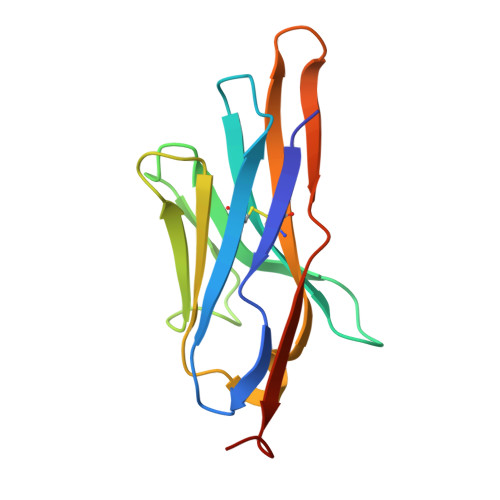
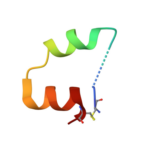
Selective molecular recognition of membrane proteins is challenging because they contain few solvent-exposed extracellular epitopes, which often depend on their native environment for structure, making them difficult to isolate faithfully for binder discovery. Here, we show that antigen-detected NMR is well suited both to characterizing the folding of engineered minimal epitopes from the human voltage-gated sodium channel Na V 1.7 and to selecting binders that recognize their solvent-exposed surfaces. Isotope labelling of the antigen enables NMR resonance assignment to assess retained local secondary structure, while 15 N titration and zz-exchange mapping provide binding and interface information. Combined with AlphaFold2 complex prediction, this creates a practical method for screening and ranking candidate binders. The approach was further validated by a high-resolution x-ray structure of an antigen-nanobody complex. Applying this workflow identified R4C8, a subtype- and species-selective nanobody whose binding to the extracellular surface of human Na V 1.7 is supported by zz-exchange mapping, modelling, and cellular recognition, and which has minimal effects on channel gating. R4C8 detected Na V 1.7 in engineered cell lines and in primary osteoarthritis-derived chondrocytes, providing a useful tool for selective target detection. These results show how antigen-detected NMR can support peptide engineering and structure-guided protein binder selection against minimal epitopes.
- Australian Institute for Bioengineering and Nanotechnology, The University of Queensland, St Lucia, Queensland, Australia.
Organizational Affiliation: