9XHH | pdb_00009xhh

Structure of the CCL19-CCR7-Gi-scFv16 complex


Experimental Data Snapshot

  • Method: ELECTRON MICROSCOPY
  • Resolution: 3.00 Å
  • Aggregation State: PARTICLE 
  • Reconstruction Method: SINGLE PARTICLE 

Starting Models: in silico, experimental
View more details

wwPDB Validation 3D Report Full Report

Validation slider image for 9XHH

This is version 1.1 of the entry. See complete history

Literature

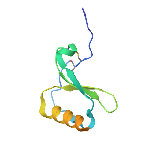
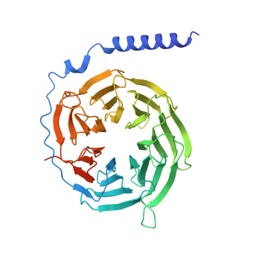
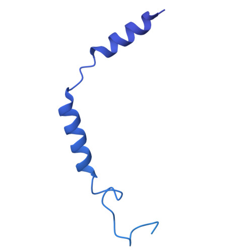
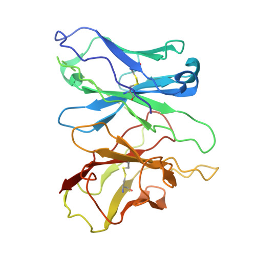
Structural insights into biased signaling at chemokine receptor CCR7.

Tanaka, K.Nishikawa, K.Shiimura, Y.Fujiyoshi, Y.Tsutsumi, N.

(2026) Proc Natl Acad Sci U S A 123: e2533975123-e2533975123

  • DOI: https://doi.org/10.1073/pnas.2533975123
  • Primary Citation Related Structures: 
    9XHH, 9XHI

  • PubMed Abstract: 

    CC chemokine receptor 7 (CCR7), which orchestrates adaptive immunity, exhibits a phenomenon known as biased agonism. CCL19 induces robust G-protein signaling and β-arrestin recruitment, leading to transient signaling. In contrast, CCL21 preferentially activates G-protein pathways with minimal arrestin engagement, resulting in sustained signaling and differential functional outcomes. Here, we present the cryo-EM structures of the human CCR7-G i complex with either CCL19 or CCL21. The structures reveal that while both engage a conserved orthosteric pocket, they adopt markedly distinct binding poses. Notably, the compact 30s loop of CCL21 inserts deeply into the receptor's extracellular vestibule, whereas the corresponding loop of CCL19 rests atop extracellular loop 2. Molecular dynamics simulations further reveal that these distinct binding modes induce differential intracellular dynamics, linked to the rotameric state of Y83 at the intracellular end of transmembrane helix 1. We demonstrate that CCL19 stabilizes a flexible conformational ensemble with a highly dynamic helix 8, creating a lateral opening favorable for GPCR kinase engagement. Conversely, CCL21 restricts this flexibility, locking the receptor in a state that precludes kinase interaction while maintaining G-protein coupling. Corroborated by functional data, these findings provide key insights into the structural basis of biased agonism at CCR7 and establish a foundation for rational design of pathway-selective immunomodulators.


  • Organizational Affiliation
    • Cellular and Structural Physiology Laboratory, Institute of Integrated Research, Institute of Science Tokyo, Bunkyo, Tokyo 113-8510, Japan.

Macromolecule Content 

  • Total Structure Weight: 236.82 kDa 
  • Atom Count: 9,463 
  • Modeled Residue Count: 1,216 
  • Deposited Residue Count: 2,134 
  • Unique protein chains: 5

Macromolecules

Find similar proteins by:|  3D Structure
Entity ID: 1
MoleculeChains  Sequence LengthOrganismDetailsImage
C-C motif chemokine 19A [auth L]92Homo sapiensMutation(s): 0 
Gene Names: CCL19ELCMIP3BSCYA19
UniProt & NIH Common Fund Data Resources
Find proteins for Q99731 (Homo sapiens)
Explore Q99731 
Go to UniProtKB:  Q99731
PHAROS:  Q99731
GTEx:  ENSG00000172724 
Entity Groups
Sequence Clusters30% Identity50% Identity70% Identity90% Identity95% Identity100% Identity
UniProt GroupQ99731
Sequence Annotations
Expand
Reference Sequence
Find similar proteins by:|  3D Structure
Entity ID: 2
MoleculeChains  Sequence LengthOrganismDetailsImage
C-C chemokine receptor type 7,Non structural polyproteinB [auth R]551Homo sapiensMutation(s): 0 
Gene Names: CCR7CMKBR7EBI1EVI1
UniProt & NIH Common Fund Data Resources
Find proteins for A0A482LYE4 (Ross River virus)
Explore A0A482LYE4 
Go to UniProtKB:  A0A482LYE4
Find proteins for P32248 (Homo sapiens)
Explore P32248 
Go to UniProtKB:  P32248
PHAROS:  P32248
GTEx:  ENSG00000126353 
Entity Groups
Sequence Clusters30% Identity50% Identity70% Identity90% Identity95% Identity100% Identity
UniProt GroupsA0A482LYE4P32248
Sequence Annotations
Expand
Reference Sequence
Find similar proteins by:|  3D Structure
Entity ID: 3
MoleculeChains  Sequence LengthOrganismDetailsImage
Guanine nucleotide-binding protein G(I)/G(S)/G(T) subunit beta-1C [auth B]371Homo sapiensMutation(s): 0 
Gene Names: GNB1
UniProt & NIH Common Fund Data Resources
Find proteins for P62873 (Homo sapiens)
Explore P62873 
Go to UniProtKB:  P62873
PHAROS:  P62873
GTEx:  ENSG00000078369 
Entity Groups
Sequence Clusters30% Identity50% Identity70% Identity90% Identity95% Identity100% Identity
UniProt GroupP62873
Sequence Annotations
Expand
Reference Sequence
Find similar proteins by:|  3D Structure
Entity ID: 4
MoleculeChains  Sequence LengthOrganismDetailsImage
Guanine nucleotide-binding protein G(I)/G(S)/G(O) subunit gamma-2,Guanine nucleotide-binding protein G(i) subunit alpha-1D [auth C],
E [auth A]
432Homo sapiensMutation(s): 0 
Gene Names: GNG2GNAI1
EC: 3.6.5
UniProt & NIH Common Fund Data Resources
Find proteins for P59768 (Homo sapiens)
Explore P59768 
Go to UniProtKB:  P59768
PHAROS:  P59768
GTEx:  ENSG00000186469 
Find proteins for P63097 (Bos taurus)
Explore P63097 
Go to UniProtKB:  P63097
Entity Groups
Sequence Clusters30% Identity50% Identity70% Identity90% Identity95% Identity100% Identity
UniProt GroupsP63097P59768
Sequence Annotations
Expand
Reference Sequence
Find similar proteins by:|  3D Structure
Entity ID: 5
MoleculeChains  Sequence LengthOrganismDetailsImage
Antibody fragment scFv16F [auth D]256Mus musculusMutation(s): 0 

Small Molecules

Ligands 1 Unique
IDChains Name / Formula / InChI Key2D Diagram3D Interactions
CLR

Query on CLR


Download:Ideal Coordinates CCD File
G [auth R]CHOLESTEROL
C27 H46 O
HVYWMOMLDIMFJA-DPAQBDIFSA-N

Experimental Data & Validation

Experimental Data

  • Method: ELECTRON MICROSCOPY
  • Resolution: 3.00 Å
  • Aggregation State: PARTICLE 
  • Reconstruction Method: SINGLE PARTICLE 
EM Software:
TaskSoftware PackageVersion
MODEL REFINEMENTPHENIX1.21.1_5286
RECONSTRUCTIONcryoSPARC4.7

Structure Validation

View Full Validation Report


Entry History 

& Funding Information

Deposition Data


Funding OrganizationLocationGrant Number
Japan Society for the Promotion of Science (JSPS)Japan24K01965
Japan Society for the Promotion of Science (JSPS)Japan24K21935
Japan Society for the Promotion of Science (JSPS)Japan22K20632

Revision History  (Full details and data files)

  • Version 1.0: 2026-04-22
    Type: Initial release
  • Version 1.1: 2026-05-06
    Changes: Data collection, Database references