Download Mendeley
Structures of the human glucose-6-phosphate transporter provide insights into its transport cycle and substrate recognition.
Zhang, W., Jiao, H., Xue, J., Zhou, J., Wang, Y., Pan, Q., Guo, Y., Zhang, G., Hu, H., Guo, X.(2026) PLoS Biol 24: e3003731-e3003731
- PubMed: 41911224 Search on PubMedSearch on PubMed Central
- DOI: https://doi.org/10.1371/journal.pbio.3003731
- Primary Citation Related Structures:
9XCA, 9XCB - PubMed Abstract:
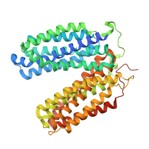
The human glucose-6-phosphate transporter (G6PT/SLC37A4) mediates the translocation of glucose-6-phosphate (G6P) from the cytoplasm into the endoplasmic reticulum, a process essential for glucose production and the maintenance of blood glucose homeostasis between meals. Dysfunction of G6PT causes glycogen storage disease type Ib (GSD-Ib), a severe metabolic disorder characterized by hypoglycemia, hepatomegaly, and neutropenia. Despite its physiological and clinical significance, the structural basis of G6P recognition and the molecular mechanisms underlying GSD-Ib have remained elusive. Here, we present cryo-electron microscopy structures of human G6PT, revealing a monomer in an outward-open state at 3.1 Å and a homodimeric assembly in a face-to-face topology at 3.3 Å. By combining computational modeling of the G6P-G6PT complexes with functional characterization, we have uncovered the key molecular elements that govern the alternating-access mechanism: an electropositive substrate-binding pocket tailored for phosphorylated sugars; conserved aromatic residues that seal the cytosolic gate; and a dynamic inter-domain salt bridge that regulates the conformational transition. Our work provides fundamental insights into the transport cycle of the organophosphate:phosphate antiporter (OPA) family, offers a framework for interpreting GSD-Ib pathology at the molecular level, and establishes a foundation for advancing the mechanistic understanding of the human SLC37 family.
- Kobilka Institute of Innovative Drug Discovery, Department of Biological Sciences, Division of Biomedical Health Sciences, School of Medicine, The Chinese University of Hong Kong, Shenzhen, Guangdong, China.
Organizational Affiliation: