9X5W | pdb_00009x5w

B/Brisbane/60/2008 HA in complex with BP-1A


Experimental Data Snapshot

  • Method: ELECTRON MICROSCOPY
  • Resolution: 2.76 Å
  • Aggregation State: PARTICLE 
  • Reconstruction Method: SINGLE PARTICLE 

Starting Model: experimental
View more details

wwPDB Validation 3D Report Full Report

Validation slider image for 9X5W

This is version 1.2 of the entry. See complete history

Literature

Broad neutralization of influenza B hemagglutinin antibodies via receptor mimicry and glycan engagement.

Huang, K.A.Nguyen, H.T.V.Chen, Y.Y.Wu, K.J.Hsu, P.H.Liu, Y.M.Lin, T.Y.Ma, C.

(2026) Proc Natl Acad Sci U S A 123: e2532989123-e2532989123

  • DOI: https://doi.org/10.1073/pnas.2532989123
  • Primary Citation Related Structures: 
    9X5W, 9X5X, 9X5Y, 9X5Z, 9X60, 9X61

  • PubMed Abstract: 

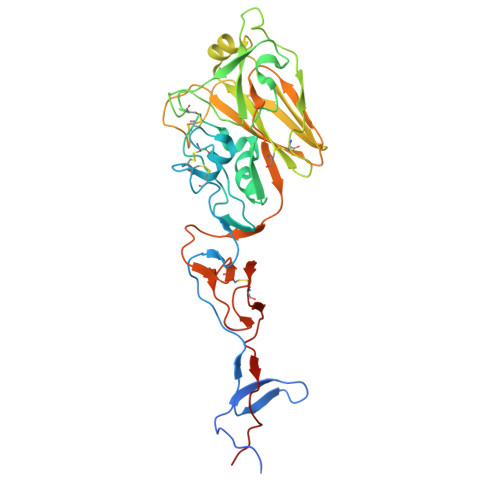
    Influenza B virus contributes to seasonal influenza epidemics and causes global morbidity and mortality. The two antigenically distinct lineages, Victoria and Yamagata, cocirculate within the population and are subject to ongoing antigenic drift. In this study, we report the isolation of cross-lineage neutralizing anti-influenza B hemagglutinin (HA) monoclonal antibodies, exhibiting hemagglutination-inhibition activities, from vaccinated adult donors. While some antibodies exhibit reduced activities against recently emerged antigenic variants, BP-1A and BO-6B demonstrate broad neutralization across influenza B viruses isolated over the past two decades and confer protection in mice against lethal challenge from both lineages. Structural analysis of the antibody Fab domains in complex with HA reveals two distinct molecular binding modes: BP-1A uses a long heavy chain CDR3 loop that mimics the ligand to target the receptor-binding site, while BO-6B engages a conserved cleft on the surface of vestigial esterase subdomain through key interactions with glycan moieties. These findings elucidate the molecular basis for broad neutralization by human anti-influenza B HA antibodies and provide insights that may guide the development of immunotherapeutics and rational vaccine design.


  • Organizational Affiliation
    • Graduate Institute of Immunology and Department of Pediatrics, National Taiwan University Hospital, College of Medicine, National Taiwan University, Taipei 100225, Taiwan.

Macromolecule Content 

  • Total Structure Weight: 433.79 kDa 
  • Atom Count: 15,978 
  • Modeled Residue Count: 2,013 
  • Deposited Residue Count: 3,912 
  • Unique protein chains: 4

Macromolecules

Find similar proteins by:|  3D Structure
Entity ID: 1
MoleculeChains  Sequence LengthOrganismDetailsImage
Hemagglutinin HA1 chain
A, C, E
362Influenza B virus (B/Brisbane/60/2008)Mutation(s): 0 
Gene Names: HA
UniProt
Find proteins for C0LT38 (Influenza B virus)
Explore C0LT38 
Go to UniProtKB:  C0LT38
Entity Groups
Sequence Clusters30% Identity50% Identity70% Identity90% Identity95% Identity100% Identity
UniProt GroupC0LT38
Glycosylation
Glycosylation Sites: 7
Sequence Annotations
Expand
Reference Sequence
Find similar proteins by:|  3D Structure
Entity ID: 2
MoleculeChains  Sequence LengthOrganismDetailsImage
Hemagglutinin HA2 chain
B, D, F
235Influenza B virus (B/Brisbane/60/2008)Mutation(s): 0 
Gene Names: HA
UniProt
Find proteins for C0LT38 (Influenza B virus)
Explore C0LT38 
Go to UniProtKB:  C0LT38
Entity Groups
Sequence Clusters30% Identity50% Identity70% Identity90% Identity95% Identity100% Identity
UniProt GroupC0LT38
Sequence Annotations
Expand
Reference Sequence
Find similar proteins by:|  3D Structure
Entity ID: 3
MoleculeChains  Sequence LengthOrganismDetailsImage
BP-1A heavy chainG [auth H],
I [auth G],
K [auth J]
474Homo sapiensMutation(s): 0 
Find similar proteins by:|  3D Structure
Entity ID: 4
MoleculeChains  Sequence LengthOrganismDetailsImage
BP-1A light chainH [auth L],
J [auth I],
L [auth K]
233Homo sapiensMutation(s): 0 

Oligosaccharides

Help  
Entity ID: 5
MoleculeChains Length2D Diagram GlycosylationD Interactions
2-acetamido-2-deoxy-beta-D-glucopyranose-(1-4)-2-acetamido-2-deoxy-beta-D-glucopyranose
M, N, O, P, Q
M, N, O, P, Q, R, S, T, U, V, W, X
2N-Glycosylation
Glycosylation Resources
GlyTouCan: G42666HT
GlyCosmos: G42666HT
GlyGen: G42666HT

Small Molecules

Ligands 1 Unique
IDChains Name / Formula / InChI Key2D Diagram3D Interactions
NAG

Query on NAG


Download:Ideal Coordinates CCD File
AA [auth A]
BA [auth C]
CA [auth C]
DA [auth C]
EA [auth E]
AA [auth A],
BA [auth C],
CA [auth C],
DA [auth C],
EA [auth E],
FA [auth E],
GA [auth E],
Y [auth A],
Z [auth A]
2-acetamido-2-deoxy-beta-D-glucopyranose
C8 H15 N O6
OVRNDRQMDRJTHS-FMDGEEDCSA-N

Experimental Data & Validation

Experimental Data

  • Method: ELECTRON MICROSCOPY
  • Resolution: 2.76 Å
  • Aggregation State: PARTICLE 
  • Reconstruction Method: SINGLE PARTICLE 
EM Software:
TaskSoftware PackageVersion
MODEL REFINEMENTPHENIX1.14_3260:
RECONSTRUCTIONcryoSPARC4.5.1

Structure Validation

View Full Validation Report


Entry History 

& Funding Information

Deposition Data


Funding OrganizationLocationGrant Number
Academia Sinica (Taiwan)TaiwanAS-IA-113-L02
Ministry of Science and Technology (MoST, Taiwan)TaiwanNSTC 114-2320-B-001 -018 -MY3

Revision History  (Full details and data files)

  • Version 1.0: 2026-04-29
    Type: Initial release
  • Version 1.1: 2026-05-13
    Changes: Data collection, Database references
  • Version 1.2: 2026-05-20
    Changes: Data collection, Database references