Download Mendeley
The phospholipid biosynthesis enzyme PlsB contains three distinct domains for membrane association, lysophosphatidic acid synthesis, and dimerization.
Li, Y., Liu, X., Li, A., Liu, Z.(2026) Protein Sci 35
- PubMed: 42324731 Search on PubMedSearch on PubMed Central
- DOI: https://doi.org/10.1002/pro.70685
- Primary Citation Related Structures:
9VUQ - PubMed Abstract:
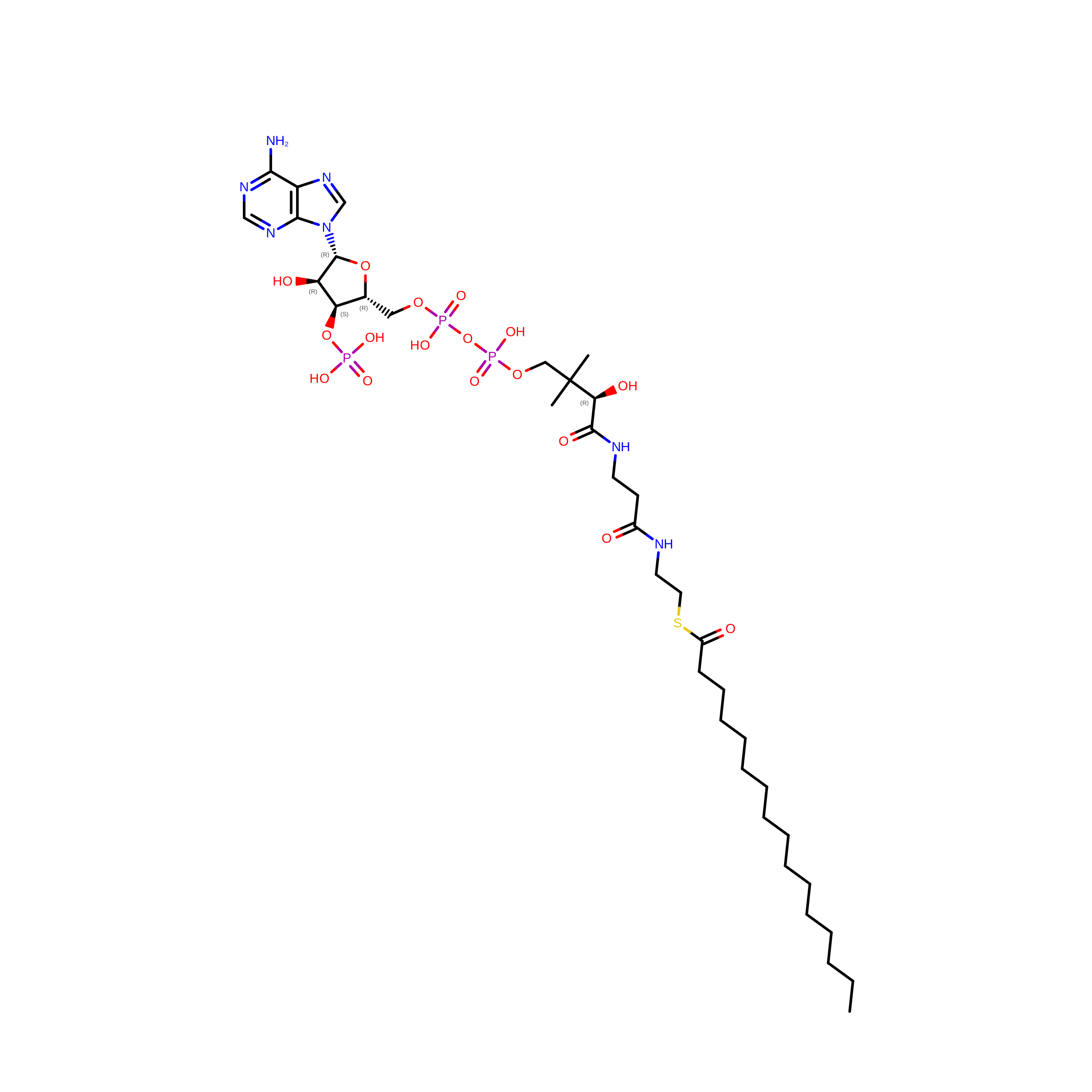
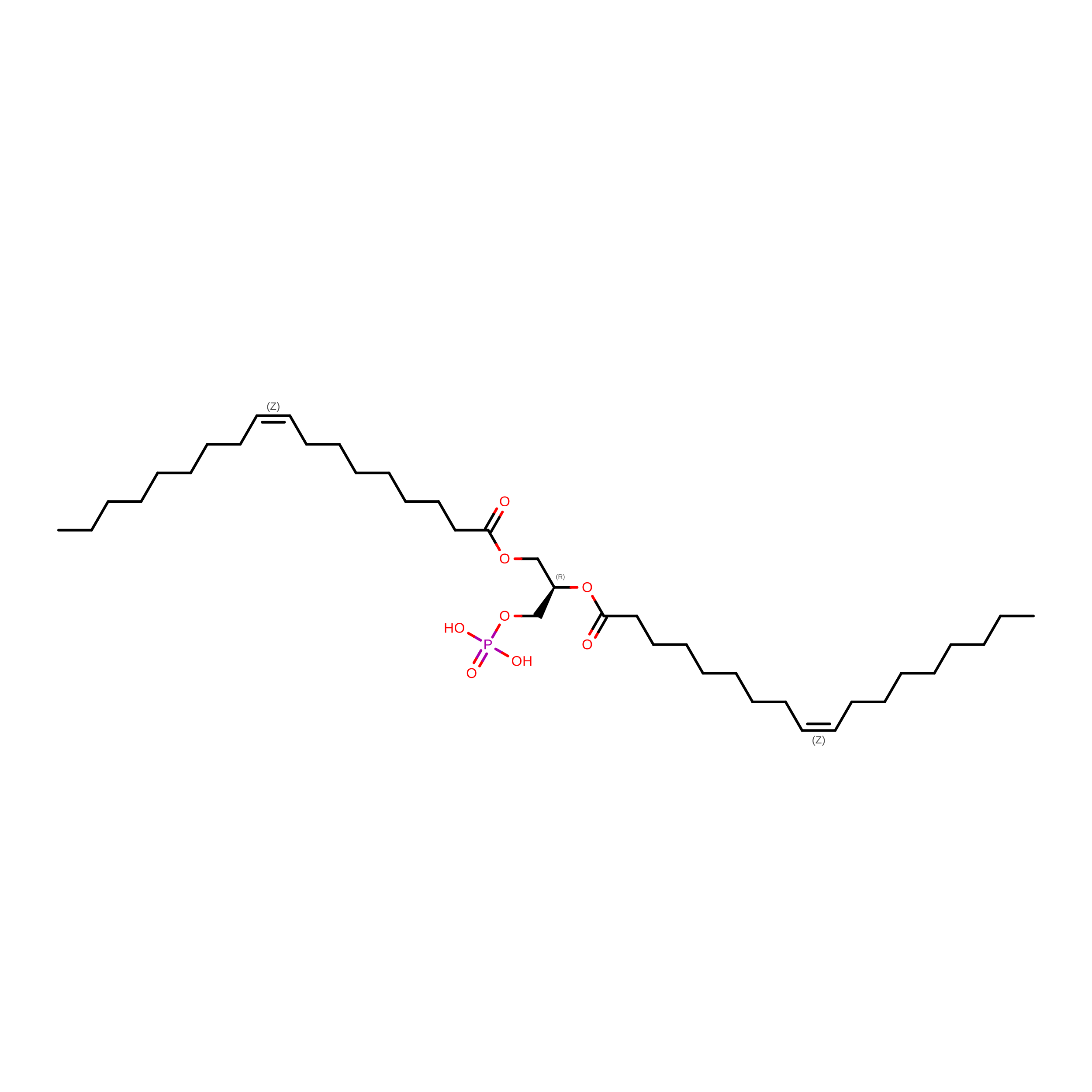
Biosynthesis of phospholipids is fundamental for membrane biogenesis in all living organisms. As a member of the glycerol-3-phosphate (G3P) acyltransferase family, PlsB is a crucial enzyme catalyzing the first step of phospholipid synthesis by converting G3P and fatty acyl-coenzyme A (CoA)/acyl carrier protein (ACP) into lysophosphatidic acid and free CoA (CoASH)/ACP. In bacterial cells, PlsB participates in the formation of antibiotic-tolerant persister cells related to multidrug tolerance, and is hence considered as a potential target for anti-persister therapy. By using the single-particle cryo-electron microscopy method, we have solved the structure of full-length PlsB from Thermomonas haemolytica (ThPlsB) at 2.79 Å resolution. The ThPlsB protein forms a homodimer with C2 symmetry and each monomer contains three distinct domains, namely the amino-terminal domain (NTD), the middle catalytic domain (MCD), and the carboxy-terminal domain. In the MCD, a fatty acyl-CoA binds in a membrane-facing surface groove enclosed by a lipid molecule 1,2-dioleoyl-sn-glycero-3-phosphate (DOPA) on one side. The interactions between ThPlsB and the membrane involve four surface-exposed amphipathic regions located in the NTD and MCD, respectively. Our structural and biochemical analysis results suggest a membrane surface association-catalysis coupling model for the PlsB-mediated biosynthesis of lysophosphatidic acid occurring at the membrane-cytosol interface.
- State Key Laboratory of Biomacromolecules, Institute of Biophysics, Chinese Academy of Sciences, Beijing, P. R. China.
Organizational Affiliation: