9VNG | pdb_00009vng

Small GTPase RhoA Y42C mutant in complex with covalent inhibitor


Experimental Data Snapshot

  • Method: X-RAY DIFFRACTION
  • Resolution: 1.43 Å
  • R-Value Free: 
    0.221 (Depositor), 0.219 (DCC) 
  • R-Value Work: 
    0.189 (Depositor), 0.190 (DCC) 
  • R-Value Observed: 
    0.191 (Depositor) 

Starting Model: experimental
View more details

wwPDB Validation 3D Report Full Report

Validation slider image for 9VNG

Ligand Structure Quality Assessment 


This is version 1.1 of the entry. See complete history

Literature

Discovering Targetable Conformation of RhoA Mutant by Integrating Native Mass Spectrometry, Ultraviolet Photodissociation, and X-ray Diffraction.

Wu, H.Liu, Z.Jiang, H.Zhao, H.Dong, C.Lu, Y.Zu, S.Guo, Y.Lai, C.Luo, P.Xu, K.Yang, Y.Yang, Y.Sun, Z.Huang, Q.Xiong, H.Zhou, L.Luo, Y.Zeng, Y.Du, D.Liang, Z.Xiao, W.Zhao, S.Zhang, W.Tang, Y.Xiao, C.Chen, K.Yang, X.Wang, F.Luo, C.

(2026) J Am Chem Soc 148: 11709-11718

  • DOI: https://doi.org/10.1021/jacs.5c20067
  • Primary Citation Related Structures: 
    8ZNY, 8ZO0, 9VNG, 9VNH, 9VNI

  • PubMed Abstract: 

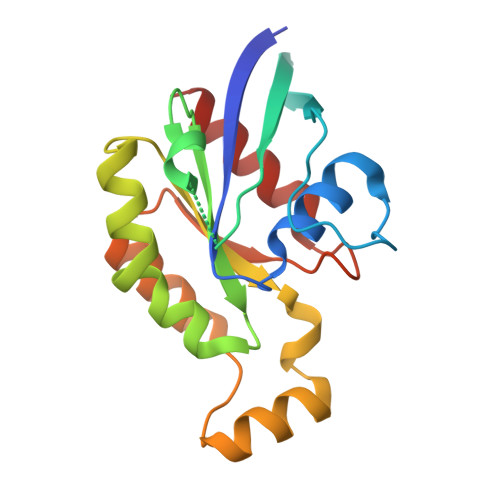
    Pathogenic mutations in "undruggable" Ras superfamily proteins challenge drug development by inducing subtle, dynamic conformational changes. Here, we integrated X-ray crystallography with native mass spectrometry and ultraviolet photodissociation (nMS-UVPD) to reveal a cryptic conformation in the oncogenic Y42C mutant of RhoA. While crystallography alone resolved two ambiguous structures, nMS-UVPD determined the dominant conformation by directly mapping the mutant's conformational dynamics, identifying an enhanced Mg 2+ -locked conformation. We explored the mechanism of mutation impairing GTP hydrolysis. This state unmasks a previously hidden, druggable pocket adjacent to Cys42, guiding our identification of a covalent inhibitor. Our integrated approach establishes a roadmap for targeting pathogenic protein mutants previously considered "undruggable" due to their highly dynamic nature.


  • Organizational Affiliation
    • State Key Laboratory of Discovery and Utilization of Functional Components in Traditional Chinese Medicine, Guizhou Medical University, Guiyang 561113, China.

Macromolecule Content 

  • Total Structure Weight: 21.12 kDa 
  • Atom Count: 1,519 
  • Modeled Residue Count: 172 
  • Deposited Residue Count: 182 
  • Unique protein chains: 1

Macromolecules

Find similar proteins by:|  3D Structure
Entity ID: 1
MoleculeChains  Sequence LengthOrganismDetailsImage
Transforming protein RhoA182Homo sapiensMutation(s): 1 
Gene Names: RHOAARH12ARHARHO12
EC: 3.6.5.2
UniProt & NIH Common Fund Data Resources
Find proteins for P61586 (Homo sapiens)
Explore P61586 
Go to UniProtKB:  P61586
PHAROS:  P61586
GTEx:  ENSG00000067560 
Entity Groups
Sequence Clusters30% Identity50% Identity70% Identity90% Identity95% Identity100% Identity
UniProt GroupP61586
Sequence Annotations
Expand
Reference Sequence

Experimental Data & Validation

Experimental Data

  • Method: X-RAY DIFFRACTION
  • Resolution: 1.43 Å
  • R-Value Free:  0.221 (Depositor), 0.219 (DCC) 
  • R-Value Work:  0.189 (Depositor), 0.190 (DCC) 
  • R-Value Observed: 0.191 (Depositor) 
Space Group: P 21 21 21
Unit Cell:
Length ( Å )Angle ( ˚ )
a = 32.04α = 90
b = 63.5β = 90
c = 82.72γ = 90
Software Package:
Software NamePurpose
PHENIXrefinement
xia2data reduction
Aimlessdata scaling
PHENIXphasing
PDB_EXTRACTdata extraction

Structure Validation

View Full Validation Report


Ligand Structure Quality Assessment 


Entry History 

& Funding Information

Deposition Data


Funding OrganizationLocationGrant Number
National Natural Science Foundation of China (NSFC)China--

Revision History  (Full details and data files)

  • Version 1.0: 2026-04-01
    Type: Initial release
  • Version 1.1: 2026-04-08
    Changes: Database references