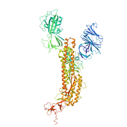
KP.3.1.1 became a dominant successor to JN.1 by the second half of 2024 but the intrinsic pathogenicity and virological feature of KP.3.1.1 remain incompletely understood. Here, we comprehensively evaluated the pathogenesis and characteristics of KP.3.1.1 in comparison to JN.1 and other JN.1-derived variants including JN.1.7, KP.2, and KP.3. The unique S31del mutation on KP.3.1.1 spike confers further evasion to the clinically authorized mAb Pemivibart and reduces convalescent serum neutralization efficiency. Structural analysis indicates that S31del induces novel glycosylation sites that facilitates evasion of neutralizing antibodies. We further reveal that S31del significantly enhances pseudovirus entry efficiency in all evaluated cell types including the human primary nasal epithelial cells. Nevertheless, the intrinsic pathogenicity of KP.3.1.1 is similar to JN.1 and KP.3, and higher than that of JN.1.7 and KP.2 in a male hamster model. Interestingly, the increased virus infectivity conferred by S31del in KP.3.1.1 spike is counterbalanced by the NSP10 S33C mutation. Overall, our study indicates that a single spike mutation can confer both enhanced immune escape and increased viral infectivity. The opposing effects of spike and non-spike mutations highlight the complex interplay of viral genomic elements in shaping their overall fitness, and reveal the high plasticity of coronavirus evolution.
Organizational Affiliation:
State Key Laboratory of Emerging Infectious Diseases, Department of Microbiology, School of Clinical Medicine, Li Ka Shing Faculty of Medicine, The University of Hong Kong, and Pandemics Research Alliance Unit at The University of Hong Kong, Hong Kong Special Administrative Region, China.
Shanghai Sci-Tech Inno Center for Infection & Immunity, National Medical Center for Infectious Diseases, Huashan Hospital, Institute of Infection and Health, Fudan University, Shanghai, China. xiaoyu_zhao@fudan.edu.cn.
Shanghai Pudong Hospital, Fudan University Pudong Medical Center, State Key Laboratory of Genetics and Development of Complex Phenotypes, MOE Engineering Research Center of Gene Technology, School of Life Sciences, Shanghai Institute of Infectious Disease and Biosecurity, Shanghai Key Laboratory of Oncology Target Discovery and Antibody Drug Development, Fudan University, Shanghai, China. xiaoyu_zhao@fudan.edu.cn.
Shanghai Fifth People's Hospital, Shanghai Institute of Infectious Disease and Biosecurity, Institutes of Biomedical Sciences, Fudan University, Shanghai, China.
Shanghai Pudong Hospital, Fudan University Pudong Medical Center, State Key Laboratory of Genetics and Development of Complex Phenotypes, MOE Engineering Research Center of Gene Technology, School of Life Sciences, Shanghai Institute of Infectious Disease and Biosecurity, Shanghai Key Laboratory of Oncology Target Discovery and Antibody Drug Development, Fudan University, Shanghai, China.
BayVax Biotech Limited, Hong Kong Science Park, Hong Kong, China.
School of Pharmaceutical Science and Technology, Hangzhou Institute for Advanced Study, University of Chinese Academy of Sciences, Hangzhou, China.
State Key Laboratory of Molecular Engineering of Polymers, Department of Macromolecular Science, Fudan University, Shanghai, China.
Department of Infectious Diseases, Nanjing Hospital of Chinese Medicine Affiliated to Nanjing University of Chinese Medicine, Nanjing, Jiangsu, China.
Xianghu Laboratory, Zhejiang, China.
School of Life and Health Technology, Dongguan University of Technology, Dongguan, China.
Centre for Virology, Vaccinology and Therapeutics, Hong Kong Science and Technology Park, Hong Kong Special Administrative Region, China.
Department of Infectious Disease and Microbiology, The University of Hong Kong-Shenzhen Hospital, Shenzhen, Guangdong Province, China.
School of Biomedical Sciences, Li Ka Shing Faculty of Medicine, The University of Hong Kong, Hong Kong Special Administrative Region, China.
Materials Innovation Institute for Life Sciences and Energy (MILES), Shenzhen, China.
Academician Workstation of Hainan Province, Hainan Medical University-The University of Hong Kong Joint Laboratory of Tropical Infectious Diseases, Haikou, Hainan Province, China.
Department of Microbiology, Queen Mary Hospital, Hong Kong Special Administrative Region, China.
Key Laboratory of Quantitative Synthetic Biology, Shenzhen Institute of Synthetic Biology, Shenzhen Institute of Advanced Technology, Chinese Academy of Sciences, Shenzhen, China.
Key Laboratory of Medical Molecular Virology (MOE/NHC/CAMS), Shanghai Institute of Infectious Disease and Biosecurity, Shanghai Frontiers Science Center of Pathogenic Microorganisms and Infection, School of Basic Medical Sciences, Shanghai Medical College, Fudan University, Shanghai, China.
School of Pharmaceutical Science and Technology, Hangzhou Institute for Advanced Study, University of Chinese Academy of Sciences, Hangzhou, China. hemaozhou@ucas.ac.cn.
Shanghai Fifth People's Hospital, Shanghai Institute of Infectious Disease and Biosecurity, Institutes of Biomedical Sciences, Fudan University, Shanghai, China. llsun@fudan.edu.cn.
Shanghai Pudong Hospital, Fudan University Pudong Medical Center, State Key Laboratory of Genetics and Development of Complex Phenotypes, MOE Engineering Research Center of Gene Technology, School of Life Sciences, Shanghai Institute of Infectious Disease and Biosecurity, Shanghai Key Laboratory of Oncology Target Discovery and Antibody Drug Development, Fudan University, Shanghai, China. pengfei_wang@fudan.edu.cn.
State Key Laboratory of Emerging Infectious Diseases, Department of Microbiology, School of Clinical Medicine, Li Ka Shing Faculty of Medicine, The University of Hong Kong, and Pandemics Research Alliance Unit at The University of Hong Kong, Hong Kong Special Administrative Region, China. hinchu@hku.hk.
Centre for Virology, Vaccinology and Therapeutics, Hong Kong Science and Technology Park, Hong Kong Special Administrative Region, China. hinchu@hku.hk.
Department of Infectious Disease and Microbiology, The University of Hong Kong-Shenzhen Hospital, Shenzhen, Guangdong Province, China. hinchu@hku.hk.
Materials Innovation Institute for Life Sciences and Energy (MILES), Shenzhen, China. hinchu@hku.hk.
AA [auth C] BA [auth C] CA [auth C] DA [auth C] EA [auth C]
AA [auth C], BA [auth C], CA [auth C], DA [auth C], EA [auth C], FA [auth C], GA [auth C], HA [auth C], J [auth A], K [auth A], L [auth A], M [auth A], N [auth A], O [auth A], P [auth A], Q [auth A], R [auth A], S [auth B], T [auth B], U [auth B], V [auth B], W [auth B], X [auth B], Y [auth B], Z [auth B]
2-acetamido-2-deoxy-beta-D-glucopyranose C8 H15 N O6 OVRNDRQMDRJTHS-FMDGEEDCSA-N