9UEP | pdb_00009uep

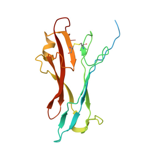
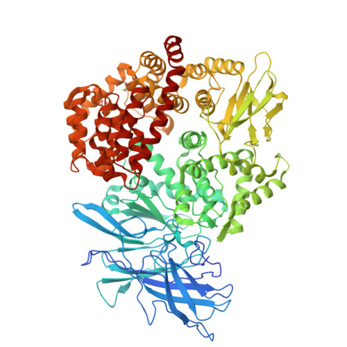
The structure of FIPV RBD and cat APN complex


Experimental Data Snapshot

  • Method: ELECTRON MICROSCOPY
  • Resolution: 2.85 Å
  • Aggregation State: PARTICLE 
  • Reconstruction Method: SINGLE PARTICLE 

wwPDB Validation 3D Report Full Report

Validation slider image for 9UEP

This is version 1.1 of the entry. See complete history

Literature

Structure and receptor recognition of type-II feline infectious peritonitis virus spike glycoprotein.

Niu, S.Tian, Y.Nguyen, L.Bai, C.Hou, X.Wang, D.Niu, Z.Liu, Z.Ren, J.Li, W.Wang, Q.Tian, W.X.Sun, J.Gao, G.F.

(2026) EMBO J 

  • DOI: https://doi.org/10.1038/s44318-026-00849-2
  • Primary Citation Related Structures: 
    9UE3, 9UEP

  • PubMed Abstract: 

    Type-II feline infectious peritonitis virus (FIPV-II) is a lethal alphacoronavirus, whose high homology with the human coronavirus CCoV-HuPn-2018 carries the risk of potential zoonotic FIPV-II transmission to humans. FIPV-II infects felids using cat aminopeptidase N (cAPN) as its cell-entry receptor, but the molecular details remain unclear. Here, we resolved cryo-electron microscopy (cryo-EM) structures of the spike (S) trimer of a representative FIPV-II strain 79-1146 (FIPV-1146), and of its complex with cAPN. The reconstructions reveal structural transitions of the S protein between the "standing" and "lying" receptor-binding domain (RBD) conformation upon complex formation with cAPN in its open conformation. Structural and mutational analyses revealed that the cAPN residues R378, D751 and R779, as well as the N748-linked glycan are essential for high-affinity RBD engagement. Cross-species analysis demonstrated narrow tropism of FIPV-1146, limited to felids and canids. Introducing the N595K and Q596R substitutions found in transmissible gastroenteritis virus (TGEV) into the FIPV-1146 RBD expands its binding capacity to APNs from pig, bovine, horse and giant panda. These findings provide insights into the entry mechanism and zoonotic constraints of FIPV-II, with potential relevance for antiviral strategies against coronaviruses.


  • Organizational Affiliation
    • Shanxi Key Laboratory of Animal Disease Research, Prevention and Control, College of Veterinary Medicine, Shanxi Agricultural University, Jinzhong, China. niusheng@sxau.edu.cn.

Macromolecule Content 

  • Total Structure Weight: 127.55 kDa 
  • Atom Count: 8,514 
  • Modeled Residue Count: 1,049 
  • Deposited Residue Count: 1,106 
  • Unique protein chains: 2

Macromolecules

Find similar proteins by:|  3D Structure
Entity ID: 1
MoleculeChains  Sequence LengthOrganismDetailsImage
Spike glycoprotein174Feline infectious peritonitis virus (strain 79-1146)Mutation(s): 0 
Gene Names: S2
UniProt
Find proteins for P10033 (Feline coronavirus (strain FIPV WSU-79/1146))
Explore P10033 
Go to UniProtKB:  P10033
Entity Groups
Sequence Clusters30% Identity50% Identity70% Identity90% Identity95% Identity100% Identity
UniProt GroupP10033
Glycosylation
Glycosylation Sites: 1
Sequence Annotations
Expand
Reference Sequence
Find similar proteins by:|  3D Structure
Entity ID: 2
MoleculeChains  Sequence LengthOrganismDetailsImage
Aminopeptidase NB [auth C]932Felis catusMutation(s): 0 
Gene Names: ANPEPAPN
EC: 3.4.11.2
UniProt
Find proteins for P79171 (Felis catus)
Explore P79171 
Go to UniProtKB:  P79171
Entity Groups
Sequence Clusters30% Identity50% Identity70% Identity90% Identity95% Identity100% Identity
UniProt GroupP79171
Glycosylation
Glycosylation Sites: 6
Sequence Annotations
Expand
Reference Sequence

Oligosaccharides

Help  
Entity ID: 3
MoleculeChains Length2D Diagram GlycosylationD Interactions
beta-D-mannopyranose-(1-4)-2-acetamido-2-deoxy-beta-D-glucopyranose-(1-4)-[alpha-L-fucopyranose-(1-6)]2-acetamido-2-deoxy-beta-D-glucopyranoseC [auth B]4N-Glycosylation
Glycosylation Resources
GlyTouCan: G32152BH
GlyCosmos: G32152BH
GlyGen: G32152BH
Entity ID: 4
MoleculeChains Length2D Diagram GlycosylationD Interactions
2-acetamido-2-deoxy-beta-D-glucopyranose-(1-4)-2-acetamido-2-deoxy-beta-D-glucopyranose
D, E
2N-Glycosylation
Glycosylation Resources
GlyTouCan: G42666HT
GlyCosmos: G42666HT
GlyGen: G42666HT

Experimental Data & Validation

Experimental Data

  • Method: ELECTRON MICROSCOPY
  • Resolution: 2.85 Å
  • Aggregation State: PARTICLE 
  • Reconstruction Method: SINGLE PARTICLE 
EM Software:
TaskSoftware PackageVersion
MODEL REFINEMENTPHENIX

Structure Validation

View Full Validation Report


Entry History 

& Funding Information

Deposition Data

  • Released Date: 2026-01-07 
  • Deposition Author(s): Sun, J.Q.

Funding OrganizationLocationGrant Number
National Natural Science Foundation of China (NSFC)China32270157

Revision History  (Full details and data files)

  • Version 1.0: 2026-01-07
    Type: Initial release
  • Version 1.1: 2026-07-22
    Changes: Data collection, Database references