Download Mendeley
Structural basis for substrate recognition in l-lysine 6-dehydrogenase from Geobacillus stearothermophilus by Cryo-EM.
Funahashi, T., Yamaguchi, H., Suzuki, S., Suzuki, H., Nishikawa, K., Takahashi, K., Tatsumi, M., Mizukoshi, T., Miyano, H., Fujiyoshi, Y., Sugiki, M.(2026) J Struct Biol 218: 108366-108366
- PubMed: 42665198 Search on PubMed
- DOI: https://doi.org/10.1016/j.jsb.2026.108366
- Primary Citation Related Structures:
9UEJ, 9UEK - PubMed Abstract:
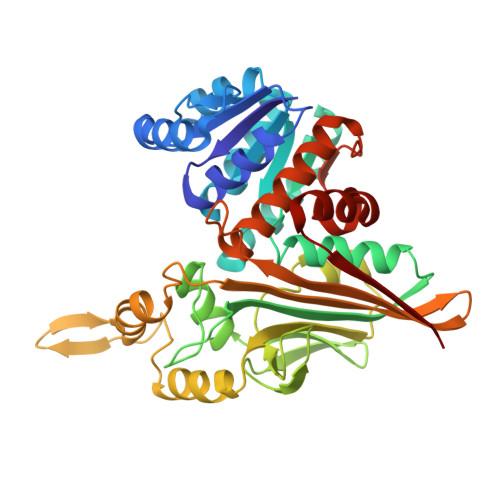
l-lysine 6-dehydrogenase (LysDH; EC 1.4.1.18) oxidatively deaminates the ε-amino group of l-lysine. Due to its high substrate specificity, LysDH serves as a valuable tool for l-lysine quantification. However, the molecular basis of this specificity has remained unclear because of the lack of substrate-bound structures. In this study, we determined the cryo-electron microscopy (cryo-EM) structures of LysDH from the thermophilic bacterium Geobacillus stearothermophilus (GstLysDH) in the apo form at 2.9 Å resolution and in complex with NAD + and l-lysine at 2.5 Å resolution. GstLysDH assembles as a tetramer, which undergoes a global conformational transition upon NAD + binding. Structural analysis revealed that the α-carboxyl and α-amino groups of l-lysine were coordinated by oppositely charged residues, thereby orienting the ε-amino group toward the nicotinamide ring of NAD + and anchoring the substrate in the optimal binding mode. This precise recognition mechanism accounts for the enzyme's strict specificity for the ε-amino group of l-lysine. Furthermore, comparative structural analysis with l-phenylalanine dehydrogenase suggests that the oxidative deamination in GstLysDH proceeds through a conserved hydride transfer mechanism. Together, these insights establish a structural framework for the rational design and industrial application of LysDH and related amino acid dehydrogenases.
- Ajinomoto Co., Inc., 1-1 Suzuki-cho, Kawasaki-ku, Kawasaki 210-8681, Japan; Institute of Integrated Research, Institute of Science Tokyo, 1-5-45 Yushima, Bunkyo-ku, Tokyo 113-8501, Japan. Electronic address: toshiya.funahashi.7ym@asv.ajinomoto.com.
Organizational Affiliation: