9UCZ | pdb_00009ucz

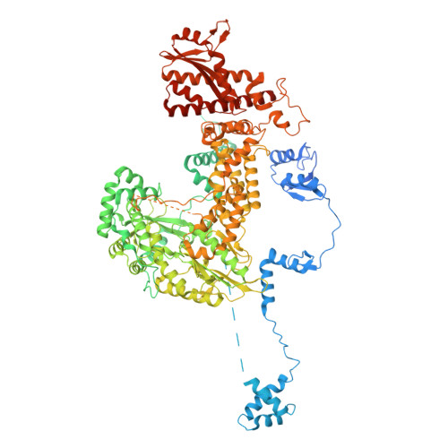
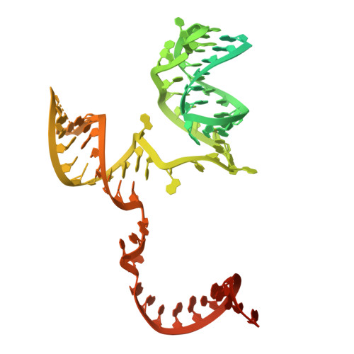
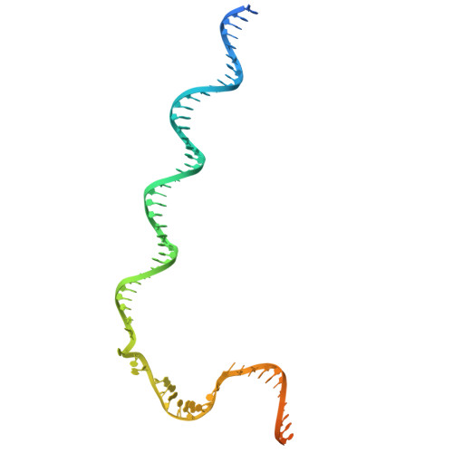
Cryo-EM structure of the R2Tg retrotransposon in complex with R2RNA and rDNA in the initiating target-primed reverse transcription state (ZF3/2 disordered)


Experimental Data Snapshot

  • Method: ELECTRON MICROSCOPY
  • Resolution: 2.61 Å
  • Aggregation State: PARTICLE 
  • Reconstruction Method: SINGLE PARTICLE 

wwPDB Validation 3D Report Full Report

Validation slider image for 9UCZ

This is version 1.1 of the entry. See complete history. 

Literature

Structural mechanism of programmable gene integration by the R2 retrotransposon coupled with CRISPR-Cas9.

Komatsu, D., Hiraizumi, M., Fell, C.W., Fujimatsu, K., Nagahata, N., Yamada, S., Sapkota, S., Ramani, H., Arya, B., Shibata, M., Yamashita, K., Gootenberg, J.S., Abudayyeh, O.O., Nishimasu, H.

(2026) Nat Struct Mol Biol 

  • DOI: https://doi.org/10.1038/s41594-026-01903-7
  • Primary Citation Related Structures: 
    9UCX, 9UCY, 9UCZ, 9UD0, 9UD1

  • PubMed Abstract: 

    R2 retroelements are non-long-terminal-repeat retrotransposons that primarily insert into 28S ribosomal DNA loci across diverse eukaryotes. R2 elements home to their target sites through target-primed reverse transcription (TPRT), in which the R2 protein recognizes and nicks the target DNA to prime reverse transcription using an R2-encoding RNA as the template. The avian R2 system from Taeniopygia guttata (R2Tg) can insert a payload sequence within an RNA template into 28S ribosomal DNA loci in human cells. Here, we present cryo-electron microscopy structures of the R2Tg-template RNA-target DNA complex in the TPRT initiation state, both alone and bound to a Cas9-guide RNA complex. These structures provide mechanistic insights into R2Tg-mediated TPRT in its natural context and in the programmable gene integration system STITCHR (site-specific target-primed insertion through targeted CRISPR homing of retroelements). Furthermore, we combine these structural insights with computational protein design methods to engineer an enhanced STITCHR system.


  • Organizational Affiliation: 
    • Department of Chemistry and Biotechnology, Graduate School of Engineering, The University of Tokyo, Tokyo, Japan.

Macromolecule Content 

  • Total Structure Weight: 234.31 kDa 
  • Atom Count: 12,119 
  • Modeled Residue Count: 1,194 
  • Deposited Residue Count: 1,565 
  • Unique protein chains: 1
  • Unique nucleic acid chains: 4

Macromolecules


Find similar proteins by:|  3D Structure
Entity ID: 1
MoleculeChains  Sequence LengthOrganismDetailsImage
Reverse transcriptase-like protein1,287Taeniopygia guttataMutation(s): 0 
Entity Groups
Sequence Clusters30% Identity50% Identity70% Identity90% Identity95% Identity100% Identity
Sequence Annotations
Expand
Reference Sequence
Find similar nucleic acids by:  (by identity cutoff) 
Entity ID: 2
MoleculeChains LengthOrganismImage
R2Tg 3'UTR RNA117Taeniopygia guttata
Sequence Annotations
Expand
Reference Sequence
Find similar nucleic acids by:  (by identity cutoff) 
Entity ID: 3
MoleculeChains LengthOrganismImage
28S DNA top strand80Taeniopygia guttata
Sequence Annotations
Expand
Reference Sequence
Find similar nucleic acids by:  (by identity cutoff) 
Entity ID: 4
MoleculeChains LengthOrganismImage
28S DNA bottom strand, 3' side50Taeniopygia guttata
Sequence Annotations
Expand
Reference Sequence
Find similar nucleic acids by:  (by identity cutoff) 
Entity ID: 5
MoleculeChains LengthOrganismImage
28S DNA bottom strand, 5' side (priming strand)31Taeniopygia guttata
Sequence Annotations
Expand
Reference Sequence

Small Molecules

Ligands 3 Unique
IDChains Name / Formula / InChI Key2D Diagram3D Interactions
TTP
(Subject of Investigation/LOI)

Query on TTP


Download:Ideal Coordinates CCD File
L [auth A]THYMIDINE-5'-TRIPHOSPHATE
C10 H17 N2 O14 P3
NHVNXKFIZYSCEB-XLPZGREQSA-N
ZN
(Subject of Investigation/LOI)

Query on ZN


Download:Ideal Coordinates CCD File
F [auth A],
G [auth A],
H [auth A],
I [auth A]
ZINC ION
Zn
PTFCDOFLOPIGGS-UHFFFAOYSA-N
MG
(Subject of Investigation/LOI)

Query on MG


Download:Ideal Coordinates CCD File
J [auth A],
K [auth A]
MAGNESIUM ION
Mg
JLVVSXFLKOJNIY-UHFFFAOYSA-N

Experimental Data & Validation

Experimental Data

  • Method: ELECTRON MICROSCOPY
  • Resolution: 2.61 Å
  • Aggregation State: PARTICLE 
  • Reconstruction Method: SINGLE PARTICLE 
EM Software:
TaskSoftware PackageVersion
RECONSTRUCTIONcryoSPARC
MODEL REFINEMENTServalcat

Structure Validation

View Full Validation Report


Entry History 

& Funding Information

Deposition Data


Funding OrganizationLocationGrant Number
Japan Science and TechnologyJapan--

Revision History  (Full details and data files)

  • Version 1.0: 2026-10-14
    Type: Initial release
  • Version 1.1: 2026-11-11
    Changes: Data collection, Database references