Biochemical characterization and X-ray structural and mutagenic analyses of the putative autolysin CdCwlT33800 catalytic domain from Clostridioides difficile.
Sekiya, H., Nonaka, Y., Kamitori, S., Tamai, E.(2025) Appl Environ Microbiol 91: e0121625-e0121625
- PubMed: 40956107 Search on PubMed
- DOI: https://doi.org/10.1128/aem.01216-25
- Primary Citation Related Structures:
9UBF - PubMed Abstract:
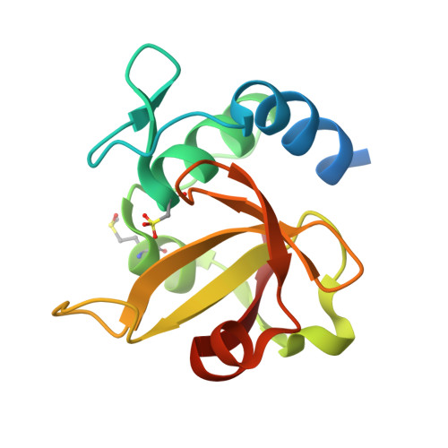
Clostridioides difficile is a major pathogen of pseudomembranous colitis, and new antimicrobial agents are needed for its treatment. Autolysins are peptidoglycan-degrading enzymes that generally reorganize the cell wall during cell division but kill bacteria by bacteriolysis when applied from outside the bacterial cell. Therefore, they have potential as novel therapeutic agents for the treatment of infectious diseases. We surveyed the genome of C. difficile strain 630 and identified two virtually identical autolysin genes, cdCwlT33800 and cdCwlT , with a lysozyme-like domain and endopeptidase domain. The entire region and each domain of the two proteins were expressed, purified, and assayed for bacteriolytic activity. Only the individual endopeptidase domain variants exhibited bacteriolytic activity against C. difficile . We also investigated the optimal pH and salt concentration, the effects of metal ions, thermostability, long-term storage, and the species specificity of the CdCwlT33800 endopeptidase domain (CdCwlT33800CD2). The structure of CdCwlT33800CD2 was elucidated by X-ray crystallography at a resolution of 1.45 Å. The overall structure was spherical and consisted of five helices and eight β-strands, with a 28 Å substrate-binding groove, at the center of which the catalytic residues of Cys242 and His296 were located. The structure of the substrate-enzyme complex was proposed through modeling and mutagenic analyses of CdCwlT33800CD2.IMPORTANCE Clostridioides difficile is a bacterium that causes severe colitis and life-threatening diarrhea, particularly after antibiotic treatment. Since current therapies are not always effective and resistance to drugs continues to increase, there is an urgent need for new treatment strategies. One promising approach is the use of lytic enzymes, which break down the bacterial cell wall and lead to bacterial death. These enzymes include autolysins, which are produced by bacteria themselves and phage-derived endolysins. In the present study, we identified a novel autolysin from C. difficile and analyzed its biochemical characteristics and structure. The present results provide insights into the development of enzyme-based therapies to combat C. difficile infections and may lead to effective alternatives to conventional antibiotics.
- Department of Infectious Disease, College of Pharmaceutical Science, Matsuyama University, Matsuyama, Ehime, Japan.
Organizational Affiliation: