Extracellular nanobody screening using conformationally stable GPCR variants.
Zhang, X., Gao, K., Nie, J., Meng, H., Sun, X., Zhao, J., Liu, X.(2025) Proc Natl Acad Sci U S A 122: e2508879122-e2508879122
- PubMed: 41187083 Search on PubMedSearch on PubMed Central
- DOI: https://doi.org/10.1073/pnas.2508879122
- Primary Citation Related Structures:
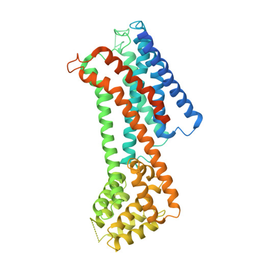
9UAP, 9UAZ, 9UCP - PubMed Abstract:
G protein-coupled receptors (GPCRs) are prominent drug targets that have attracted intensive efforts in drug screening. Binding-based screening methods for GPCR ligands often require conformationally stable, purified receptors. However, obtaining large quantities of GPCRs in stable states, particularly with unoccupied extracellular ligand-binding pockets and especially in their active conformations, remains challenging due to the inherent dynamic nature of these receptors. To address this challenge, we propose a universal approach for stabilizing GPCRs in specific conformations. Using the M1 muscarinic acetylcholine receptor (M1R) as a model, we successfully stabilized M1R in its active conformation through de novo design of a fusion protein, and further demonstrated the generalizability of this strategy by applying it to other GPCRs. We screened a synthetic yeast display library of nanobodies against both the stabilized active-state and previously reported inactive-state M1R, identifying several nanobodies that specifically recognize each conformation. This method not only facilitates the stabilization of GPCRs in desired states but also provides valuable tools for developing more selective therapeutic agents, enhancing drug discovery efficiency and specificity.
- State Key Laboratory of Membrane Biology, Tsinghua-Peking Center for Life Sciences, School of Pharmaceutical Sciences, Tsinghua University, Beijing 100084, China.
Organizational Affiliation: