9UAJ | pdb_00009uaj

Ovorubin from the golden apple snail (Pomacea canaliculata)


Experimental Data Snapshot

  • Method: ELECTRON MICROSCOPY
  • Resolution: 2.12 Å
  • Aggregation State: PARTICLE 
  • Reconstruction Method: SINGLE PARTICLE 

Starting Models: other
View more details

wwPDB Validation 3D Report Full Report

Validation slider image for 9UAJ

This is version 1.0 of the entry. See complete history

Literature

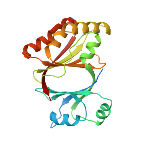
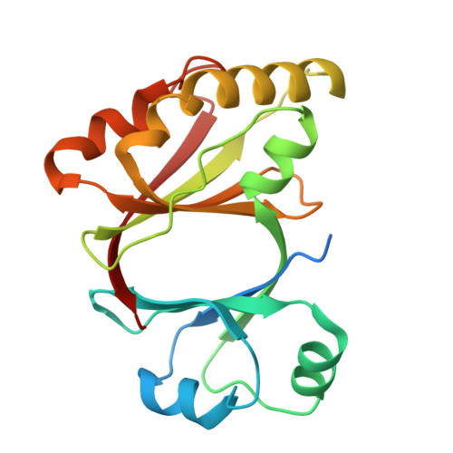
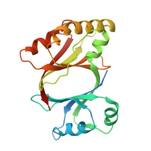
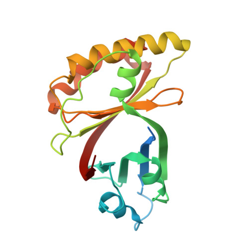
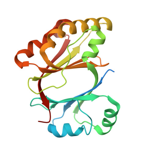
Structure of the chromoprotein ovorubin from the golden apple snail (Pomacea canaliculata).

Wilasluck, P.Saw, W.G.Tran, B.N.Sabat, G.Shih, O.Hengphasatporn, K.Shigeta, Y.Vinayavekhin, N.Wangkanont, K.

(2026) Protein Sci 35: e70439-e70439

  • DOI: https://doi.org/10.1002/pro.70439
  • Primary Citation Related Structures: 
    9UAJ

  • PubMed Abstract: 

    The golden apple snail (Pomacea canaliculata), an invasive gastropod, produces distinctly bright pink egg masses. The astaxanthin-binding ovorubin (P. canaliculata ovorubin [PcOvo]) is a 300-kDa glycoprotein responsible for the egg coloration. Here, we determine the three-dimensional structure of PcOvo using cryo-electron microscopy (cryo-EM). PcOvo is a heterodecameric protein consisting of two copies of each PcOvo1-5 subunit. The subunits have similar repeated ferredoxin-like structures. N-linked glycosylation sites are identified. Solution x-ray scattering data support the overall architecture of the complex. PcOvo3 and PcOvo5 have hydrophobic pockets that likely bind various hydrophobic compounds. The cryo-EM map and binding experiments suggest that PcOvo5 binds astaxanthin, which is responsible for the pink color of the protein. Our structure provides molecular insights into the nature of the gastropod egg coloration and lays a foundation for further investigation of ovorubin biology and evolution.


  • Organizational Affiliation
    • Center of Excellence for Molecular Biology and Genomics of Shrimp, Department of Biochemistry, Faculty of Science, Chulalongkorn University, Bangkok, Thailand.

Macromolecule Content 

  • Total Structure Weight: 234.22 kDa 
  • Atom Count: 15,208 
  • Modeled Residue Count: 1,817 
  • Deposited Residue Count: 2,032 
  • Unique protein chains: 5

Macromolecules

Find similar proteins by:|  3D Structure
Entity ID: 1
MoleculeChains  Sequence LengthOrganismDetailsImage
PcOvo5
A, F
204Pomacea canaliculataMutation(s): 0 
UniProt
Find proteins for A0A2T7NVP5 (Pomacea canaliculata)
Explore A0A2T7NVP5 
Go to UniProtKB:  A0A2T7NVP5
Entity Groups
Sequence Clusters30% Identity50% Identity70% Identity90% Identity95% Identity100% Identity
UniProt GroupA0A2T7NVP5
Glycosylation
Glycosylation Sites: 1
Sequence Annotations
Expand
Reference Sequence
Find similar proteins by:|  3D Structure
Entity ID: 2
MoleculeChains  Sequence LengthOrganismDetailsImage
PcOvo3s
B, J
199Pomacea canaliculataMutation(s): 0 
UniProt
Find proteins for A0A2T7NVP5 (Pomacea canaliculata)
Explore A0A2T7NVP5 
Go to UniProtKB:  A0A2T7NVP5
Entity Groups
Sequence Clusters30% Identity50% Identity70% Identity90% Identity95% Identity100% Identity
UniProt GroupA0A2T7NVP5
Glycosylation
Glycosylation Sites: 1
Sequence Annotations
Expand
Reference Sequence
Find similar proteins by:|  3D Structure
Entity ID: 3
MoleculeChains  Sequence LengthOrganismDetailsImage
Perivitellin ovorubin-1
C, I
200Pomacea canaliculataMutation(s): 0 
UniProt
Find proteins for J7I2T6 (Pomacea canaliculata)
Explore J7I2T6 
Go to UniProtKB:  J7I2T6
Entity Groups
Sequence Clusters30% Identity50% Identity70% Identity90% Identity95% Identity100% Identity
UniProt GroupJ7I2T6
Sequence Annotations
Expand
Reference Sequence
Find similar proteins by:|  3D Structure
Entity ID: 4
MoleculeChains  Sequence LengthOrganismDetailsImage
Perivitellin protein
D, H
203Pomacea canaliculataMutation(s): 0 
UniProt
Find proteins for A0A2T7NVP6 (Pomacea canaliculata)
Explore A0A2T7NVP6 
Go to UniProtKB:  A0A2T7NVP6
Entity Groups
Sequence Clusters30% Identity50% Identity70% Identity90% Identity95% Identity100% Identity
UniProt GroupA0A2T7NVP6
Glycosylation
Glycosylation Sites: 2
Sequence Annotations
Expand
Reference Sequence
Find similar proteins by:|  3D Structure
Entity ID: 5
MoleculeChains  Sequence LengthOrganismDetailsImage
Perivitellin ovorubin-2
E, G
210Pomacea canaliculataMutation(s): 0 
UniProt
Find proteins for J7HZ90 (Pomacea canaliculata)
Explore J7HZ90 
Go to UniProtKB:  J7HZ90
Entity Groups
Sequence Clusters30% Identity50% Identity70% Identity90% Identity95% Identity100% Identity
UniProt GroupJ7HZ90
Glycosylation
Glycosylation Sites: 2
Sequence Annotations
Expand
Reference Sequence

Oligosaccharides

Help  
Entity ID: 6
MoleculeChains Length2D Diagram GlycosylationD Interactions
alpha-L-fucopyranose-(1-3)-[2-acetamido-2-deoxy-beta-D-glucopyranose-(1-4)]2-acetamido-2-deoxy-beta-D-glucopyranose
K, O, P, R
3N-Glycosylation
Glycosylation Resources
GlyTouCan: G11971MR
GlyCosmos: G11971MR
GlyGen: G11971MR
Entity ID: 7
MoleculeChains Length2D Diagram GlycosylationD Interactions
2-acetamido-2-deoxy-beta-D-glucopyranose-(1-4)-2-acetamido-2-deoxy-beta-D-glucopyranose
L, M, N, Q, S
L, M, N, Q, S, T
2N-Glycosylation
Glycosylation Resources
GlyTouCan: G42666HT
GlyCosmos: G42666HT
GlyGen: G42666HT

Experimental Data & Validation

Experimental Data

  • Method: ELECTRON MICROSCOPY
  • Resolution: 2.12 Å
  • Aggregation State: PARTICLE 
  • Reconstruction Method: SINGLE PARTICLE 
EM Software:
TaskSoftware PackageVersion
RECONSTRUCTIONcryoSPARC4.6.0
MODEL REFINEMENTPHENIX1.21.2_5419

Structure Validation

View Full Validation Report


Entry History 

& Funding Information

Deposition Data


Funding OrganizationLocationGrant Number
Other governmentThailandChulalongkorn University

Revision History  (Full details and data files)

  • Version 1.0: 2025-12-31
    Type: Initial release