


Serine Beta-Lactamase OXA-48 in complex with Dual MBL/SBL inhibitor FB1-9
Li, G.-B., Liang, G.-Q.To be published.
Experimental Data Snapshot
Starting Model: experimental
View more details
Macromolecule Content
Entity ID: 1 | |||||
|---|---|---|---|---|---|
| Molecule | Chains | Sequence Length | Organism | Details | Image |
| Beta-lactamase OXA-48 | 242 | Klebsiella pneumoniae | Mutation(s): 0 Gene Names: OXA-48, bla OXA-48, bla_4, blaOXA-48, KPE71T_00045, SAMEA3727706_05517 EC: 3.5.2.6 | 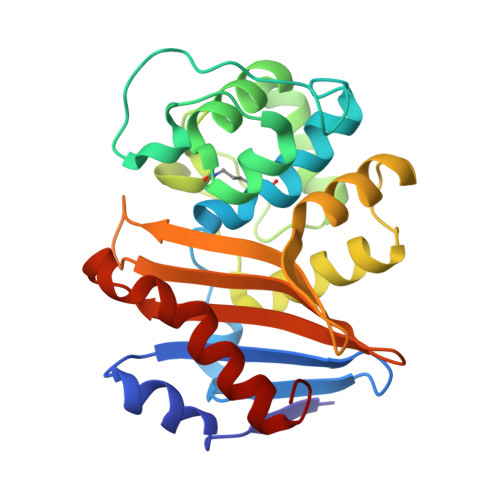 | |
UniProt | |||||
Entity Groups | |||||
| Sequence Clusters | 30% Identity50% Identity70% Identity90% Identity95% Identity100% Identity | ||||
| UniProt Group | Q6XEC0 | ||||
Sequence AnnotationsExpand | |||||
Reference Sequence | |||||
| Ligands 1 Unique | |||||
|---|---|---|---|---|---|
| ID | Chains | Name / Formula / InChI Key | 2D Diagram | 3D Interactions | |
| A1EOK (Subject of Investigation/LOI) Download:Ideal Coordinates CCD File | I [auth A] J [auth B] K [auth C] L [auth D] M [auth E] | 7-[1-(2-fluoranylethyl)azetidin-3-yl]oxy-2-oxidanyl-3,4-dihydro-1,2-benzoxaborinine-8-carboxylic acid C14 H17 B F N O5 MWTHZUDXLDLHKV-UHFFFAOYSA-N |  | ||
| Modified Residues 1 Unique | |||||
|---|---|---|---|---|---|
| ID | Chains | Type | Formula | 2D Diagram | Parent |
| KCX Query on KCX | A, B, C, D, E A, B, C, D, E, F, G, H | L-PEPTIDE LINKING | C7 H14 N2 O4 |  | LYS |
| Length ( Å ) | Angle ( ˚ ) |
|---|---|
| a = 58.419 | α = 89.86 |
| b = 93.992 | β = 90.06 |
| c = 106.226 | γ = 107.81 |
| Software Name | Purpose |
|---|---|
| PHENIX | refinement |
| SCALA | data scaling |
| PHASER | phasing |
| HKL-3000 | data reduction |
| Funding Organization | Location | Grant Number |
|---|---|---|
| National Natural Science Foundation of China (NSFC) | China | -- |


