9U98 | pdb_00009u98

Glycoside hydrolase family 1 beta-glucosidase (E318G mutant) from Streptomyces griseus (sophorose complex)


Experimental Data Snapshot

  • Method: X-RAY DIFFRACTION
  • Resolution: 2.13 Å
  • R-Value Free: 
    0.228 (Depositor), 0.233 (DCC) 
  • R-Value Work: 
    0.181 (Depositor), 0.190 (DCC) 

Starting Model: experimental
View more details

wwPDB Validation 3D Report Full Report

Validation slider image for 9U98

Ligand Structure Quality Assessment 


This is version 1.1 of the entry. See complete history

Literature

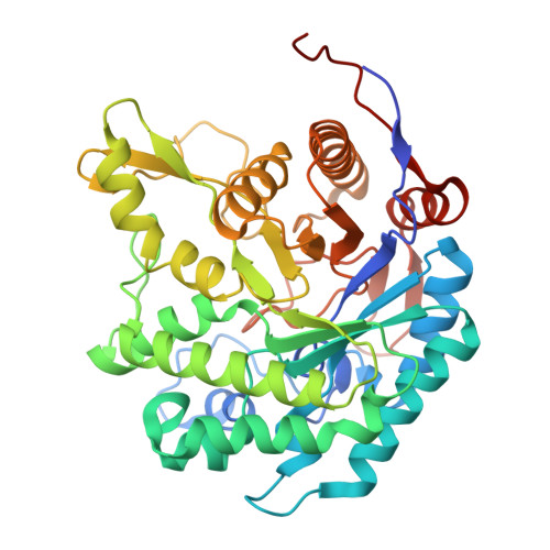
A beta-1,2-glucan-associated glycoside hydrolase family 1 beta-glucosidase from Streptomyces griseus.

Kumakura, H.Motouchi, S.Kobayashi, K.Inoue, M.Kariuda, N.Nakai, H.Nakajima, M.

(2025) Protein Sci 34: e70255-e70255

  • DOI: https://doi.org/10.1002/pro.70255
  • Primary Citation Related Structures: 
    8KAP, 9U98

  • PubMed Abstract: 

    β-Glucosidases, major enzymes that release glucose from various natural compounds, are phylogenetically classified into glycoside hydrolase (GH) families. GH1 is the largest of these families. No β-1,2-glucan-associated GH1 enzyme has been found, even though β-1,2-glucans are natural carbohydrates that are important for interaction between organisms and environmental adaptation. In this study, functional and structural analyses of a GH1 enzyme from Streptomyces griseus (SGR_2426 protein) were performed. SGR_2426 showed the highest hydrolytic activity toward p-nitrophenyl β-glucopyranoside among p-nitrophenyl sugars. This enzyme showed hydrolytic activity toward β-1,2-glucooligosaccharides specifically among β-linked glucooligosaccharides. A structure of the enzyme in complex with sophorose (β-1,2-glucodisaccharide) was obtained as a Michaelis complex. The six-membered ring of the glucose unit at the reducing end of sophorose is positioned in a hydrophobic environment between Trp291 and Met171, while only residue Gln229 forms a hydrogen bond directly. Trp291 and Gln229 are proposed as candidates for the residues important for substrate specificity based on comparison with structurally characterized GH1 homologs. Mutational analysis of Trp291 and Gln229 suggested that Trp291 is important for substrate recognition but not for substrate specificity and that Gln229 is involved in substrate specificity. SGR_2426 is the first identified β-1,2-glucan-associated β-glucosidase in the GH1 family.


  • Organizational Affiliation
    • Department of Applied Biological Science, Faculty of Science and Technology, Tokyo University of Science, Noda, Japan.

Macromolecule Content 

  • Total Structure Weight: 185.28 kDa 
  • Atom Count: 12,837 
  • Modeled Residue Count: 1,560 
  • Deposited Residue Count: 1,656 
  • Unique protein chains: 1

Macromolecules

Find similar proteins by:|  3D Structure
Entity ID: 1
MoleculeChains  Sequence LengthOrganismDetailsImage
Putative beta-glucosidase
A, B, C, D
414Streptomyces griseus subsp. griseus NBRC 13350Mutation(s): 1 
Gene Names: SGR_2426
UniProt
Find proteins for B1W1N0 (Streptomyces griseus subsp. griseus (strain JCM 4626 / CBS 651.72 / NBRC 13350 / KCC S-0626 / ISP 5235))
Explore B1W1N0 
Go to UniProtKB:  B1W1N0
Entity Groups
Sequence Clusters30% Identity50% Identity70% Identity90% Identity95% Identity100% Identity
UniProt GroupB1W1N0
Sequence Annotations
Expand
Reference Sequence

Oligosaccharides

Help  
Entity ID: 2
MoleculeChains Length2D Diagram GlycosylationD Interactions
beta-D-glucopyranose-(1-2)-beta-D-glucopyranose
E, H
2N/A
Glycosylation Resources
GlyTouCan: G26080VZ
GlyCosmos: G26080VZ
Entity ID: 3
MoleculeChains Length2D Diagram GlycosylationD Interactions
beta-D-glucopyranose-(1-2)-alpha-D-glucopyranose
F, G, I
2N/A
Glycosylation Resources
GlyTouCan: G71571NA
GlyCosmos: G71571NA

Small Molecules

Ligands 3 Unique
IDChains Name / Formula / InChI Key2D Diagram3D Interactions
BGC
(Subject of Investigation/LOI)

Query on BGC


Download:Ideal Coordinates CCD File
R [auth D]beta-D-glucopyranose
C6 H12 O6
WQZGKKKJIJFFOK-VFUOTHLCSA-N
PGE
(Subject of Investigation/LOI)

Query on PGE


Download:Ideal Coordinates CCD File
J [auth A],
M [auth B],
P [auth C]
TRIETHYLENE GLYCOL
C6 H14 O4
ZIBGPFATKBEMQZ-UHFFFAOYSA-N
CL
(Subject of Investigation/LOI)

Query on CL


Download:Ideal Coordinates CCD File
K [auth A],
L [auth A],
N [auth B],
O [auth B],
Q [auth C]
CHLORIDE ION
Cl
VEXZGXHMUGYJMC-UHFFFAOYSA-M

Experimental Data & Validation

Experimental Data

  • Method: X-RAY DIFFRACTION
  • Resolution: 2.13 Å
  • R-Value Free:  0.228 (Depositor), 0.233 (DCC) 
  • R-Value Work:  0.181 (Depositor), 0.190 (DCC) 
Space Group: P 1 21 1
Unit Cell:
Length ( Å )Angle ( ˚ )
a = 91.793α = 90
b = 100.179β = 102.868
c = 96.048γ = 90
Software Package:
Software NamePurpose
REFMACrefinement
XDSdata reduction
Aimlessdata scaling
MOLREPphasing
ARP/wARPmodel building

Structure Validation

View Full Validation Report


Ligand Structure Quality Assessment 


Entry History 

& Funding Information

Deposition Data


Funding OrganizationLocationGrant Number
Japan Society for the Promotion of Science (JSPS)Japan20K05830
Japan Science and TechnologyJapanJPMJSP2151

Revision History  (Full details and data files)

  • Version 1.0: 2025-04-16
    Type: Initial release
  • Version 1.1: 2025-10-29
    Changes: Database references