


Complex structure of a glycosyltransferase
Yu, H.J., Zhang, M., Sun, H.H., Liu, X.T.To be published.
Experimental Data Snapshot
wwPDB Validation 3D Report Full Report

Entity ID: 1 | |||||
|---|---|---|---|---|---|
| Molecule | Chains | Sequence Length | Organism | Details | Image |
| Procollagen galactosyltransferase 1 | 613 | Homo sapiens | Mutation(s): 0 Gene Names: COLGALT1, GLT25D1, PSEC0241 EC: 2.4.1.50 | 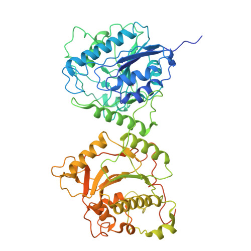 | |
UniProt & NIH Common Fund Data Resources | |||||
PHAROS: Q8NBJ5 GTEx: ENSG00000130309 | |||||
Entity Groups | |||||
| Sequence Clusters | 30% Identity50% Identity70% Identity90% Identity95% Identity100% Identity | ||||
| UniProt Group | Q8NBJ5 | ||||
Sequence AnnotationsExpand | |||||
Reference Sequence | |||||
Entity ID: 2 | |||||
|---|---|---|---|---|---|
| Molecule | Chains | Sequence Length | Organism | Details | Image |
| SER-GLY-ALA-LYZ-GLY-GLU-LYS-GLY-SER | B [auth C] | 9 | Homo sapiens | Mutation(s): 0 | 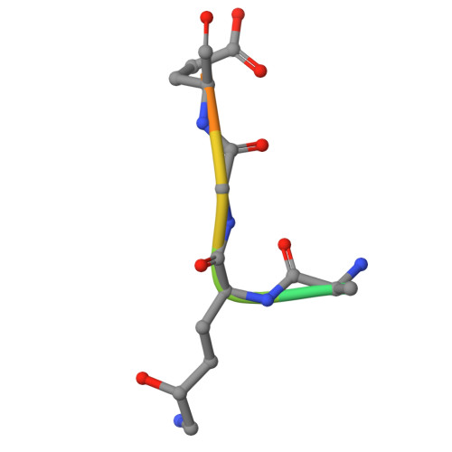 |
| Ligands 3 Unique | |||||
|---|---|---|---|---|---|
| ID | Chains | Name / Formula / InChI Key | 2D Diagram | 3D Interactions | |
| GDU (Subject of Investigation/LOI) Download:Ideal Coordinates CCD File | C [auth A] | GALACTOSE-URIDINE-5'-DIPHOSPHATE C15 H24 N2 O17 P2 HSCJRCZFDFQWRP-ABVWGUQPSA-N |  | ||
| UDP (Subject of Investigation/LOI) Download:Ideal Coordinates CCD File | E [auth A] | URIDINE-5'-DIPHOSPHATE C9 H14 N2 O12 P2 XCCTYIAWTASOJW-XVFCMESISA-N |  | ||
| MN (Subject of Investigation/LOI) Download:Ideal Coordinates CCD File | D [auth A], F [auth A] | MANGANESE (II) ION Mn WAEMQWOKJMHJLA-UHFFFAOYSA-N |  | ||
| Modified Residues 1 Unique | |||||
|---|---|---|---|---|---|
| ID | Chains | Type | Formula | 2D Diagram | Parent |
| LYZ Query on LYZ | B [auth C] | L-PEPTIDE LINKING | C6 H14 N2 O3 |  | LYS |
| Task | Software Package | Version |
|---|---|---|
| MODEL REFINEMENT | PHENIX | |
| RECONSTRUCTION | RELION | |
| RECONSTRUCTION | RELION | |
| RECONSTRUCTION | RELION |
| Funding Organization | Location | Grant Number |
|---|---|---|
| National Natural Science Foundation of China (NSFC) | China | -- |


