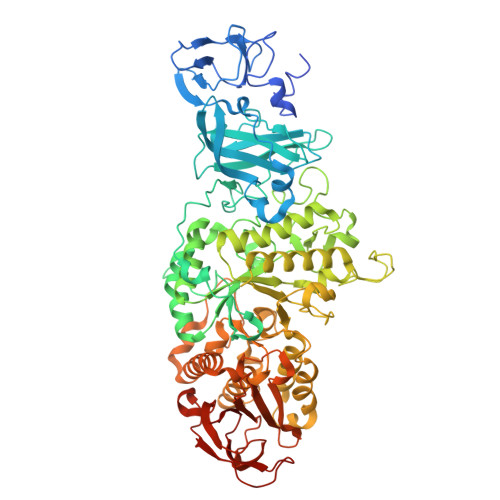
9U77 | pdb_00009u77

Crystal structure of Glycogen branching enzyme (VvGBE) from Vibrio vulnificus MO6-24/O


Experimental Data Snapshot

  • Method: X-RAY DIFFRACTION
  • Resolution: 2.50 Å
  • R-Value Free: 
    0.257 (Depositor), 0.260 (DCC) 
  • R-Value Work: 
    0.216 (Depositor), 0.217 (DCC) 
  • R-Value Observed: 
    0.217 (Depositor) 

Starting Model: experimental
View more details

wwPDB Validation 3D Report Full Report

Validation slider image for 9U77

This is version 1.1 of the entry. See complete history

Literature

Structural and functional analysis of transglycosylation mechanism of glycogen branching enzyme from Vibrio vulnificus.

An, Y.Tran, P.L.Lee, S.J.Ahn, W.C.Park, K.H.Woo, E.J.Park, J.T.

(2026) Int J Biol Macromol 343: 150415-150415

  • DOI: https://doi.org/10.1016/j.ijbiomac.2026.150415
  • Primary Citation Related Structures: 
    9U77

  • PubMed Abstract: 

    Glycogen branching enzymes (GBEs) catalyze the formation of α-1,6-glucosidic branches during glycogen biosynthesis. In this study, we characterized the GBE from Vibrio vulnificus (VvGBE), highlighting its distinct transglycosylation activity and capacity to generate short-chain branches from defined maltooligosaccharides. The VvGBE crystal structure revealed a typical (β/α) 8 -barrel fold comprising four domains (N1, N2, A, and C), characteristic of type I GBEs. Key residues, including Y229, F312, N389, D423, and E476, formed a negatively charged substrate-binding pocket, with N389 influencing branch length by favoring shorter chains. Biochemical assays showed that VvGBE preferentially transfers glucan chains with a peak degree of polymerization (DP) of 5. Structural superposition with the cceBE1 ligand model indicated that its binding pocket accommodates six glucose residues, with Y229 playing a role in determining the branching pattern. Substrate analysis revealed that a minimum DP of 11 is required for branch activity. Shorter substrates (< 11 glucose units) bound at the A1 site (M490, F489) for α-1,4-chain elongation, whereas longer substrates (≥11 units) adopt a U-shaped conformation at the A2 site (W644, V530) to form α-1,6-branches, with Y433 essential for proper acceptor positioning. These results refine our understanding of GBE substrate specificity and provide a structural framework for enzyme engineering.


  • Organizational Affiliation
    • Jilin Medical University, Jilin, 132013, People's Republic of China.

Macromolecule Content 

  • Total Structure Weight: 170.28 kDa 
  • Atom Count: 11,896 
  • Modeled Residue Count: 1,434 
  • Deposited Residue Count: 1,484 
  • Unique protein chains: 1

Macromolecules

Find similar proteins by:|  3D Structure
Entity ID: 1
MoleculeChains  Sequence LengthOrganismDetailsImage
1,4-alpha-glucan branching enzyme GlgB
A, B
742Vibrio vulnificus MO6-24/OMutation(s): 0 
Gene Names: glgBCRN52_00655
EC: 2.4.1.18
UniProt
Find proteins for A0A2S3R8T2 (Vibrio vulnificus)
Explore A0A2S3R8T2 
Go to UniProtKB:  A0A2S3R8T2
Entity Groups
Sequence Clusters30% Identity50% Identity70% Identity90% Identity95% Identity100% Identity
UniProt GroupA0A2S3R8T2
Sequence Annotations
Expand
Reference Sequence

Experimental Data & Validation

Experimental Data

  • Method: X-RAY DIFFRACTION
  • Resolution: 2.50 Å
  • R-Value Free:  0.257 (Depositor), 0.260 (DCC) 
  • R-Value Work:  0.216 (Depositor), 0.217 (DCC) 
  • R-Value Observed: 0.217 (Depositor) 
Space Group: C 1 2 1
Unit Cell:
Length ( Å )Angle ( ˚ )
a = 166.8α = 90
b = 106.063β = 123.3
c = 141.126γ = 90
Software Package:
Software NamePurpose
PHENIXrefinement
HKL-2000data reduction
HKL-2000data scaling
PHENIXphasing

Structure Validation

View Full Validation Report


Entry History 

& Funding Information

Deposition Data


Funding OrganizationLocationGrant Number
Not funded--

Revision History  (Full details and data files)

  • Version 1.0: 2026-02-04
    Type: Initial release
  • Version 1.1: 2026-08-26
    Changes: Database references