


Crystal structure of human glutaminyl cyclase in complex with Inhibitor CL21
Li, G.-B., Meng, F.-B., Chen, Y.-T.To be published.
Experimental Data Snapshot
Starting Model: experimental
View more details
Entity ID: 1 | |||||
|---|---|---|---|---|---|
| Molecule | Chains | Sequence Length | Organism | Details | Image |
| Glutaminyl-peptide cyclotransferase | 361 | Homo sapiens | Mutation(s): 0 Gene Names: QPCT EC: 2.3.2.5 | 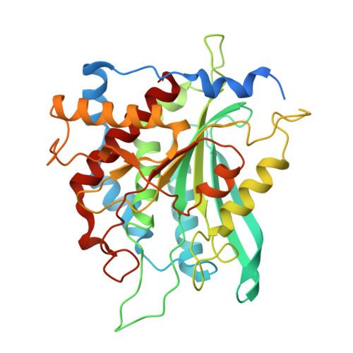 | |
UniProt & NIH Common Fund Data Resources | |||||
PHAROS: Q16769 GTEx: ENSG00000115828 | |||||
Entity Groups | |||||
| Sequence Clusters | 30% Identity50% Identity70% Identity90% Identity95% Identity100% Identity | ||||
| UniProt Group | Q16769 | ||||
Sequence AnnotationsExpand | |||||
Reference Sequence | |||||
| Ligands 4 Unique | |||||
|---|---|---|---|---|---|
| ID | Chains | Name / Formula / InChI Key | 2D Diagram | 3D Interactions | |
| A1EN4 (Subject of Investigation/LOI) Download:Ideal Coordinates CCD File | B [auth A] | 3-(2,4-dimethoxyphenyl)-6-[(5-methylimidazol-1-yl)methyl]-1H-pyridin-2-one C18 H19 N3 O3 ZECHXPHSMKLWSM-UHFFFAOYSA-N |  | ||
| GOL Download:Ideal Coordinates CCD File | G [auth A], H [auth A], I [auth A], J [auth A] | GLYCEROL C3 H8 O3 PEDCQBHIVMGVHV-UHFFFAOYSA-N |  | ||
| ZN Download:Ideal Coordinates CCD File | C [auth A] | ZINC ION Zn PTFCDOFLOPIGGS-UHFFFAOYSA-N |  | ||
| MG Download:Ideal Coordinates CCD File | D [auth A], E [auth A], F [auth A] | MAGNESIUM ION Mg JLVVSXFLKOJNIY-UHFFFAOYSA-N |  | ||
| Length ( Å ) | Angle ( ˚ ) |
|---|---|
| a = 277.712 | α = 90 |
| b = 277.712 | β = 90 |
| c = 277.712 | γ = 90 |
| Software Name | Purpose |
|---|---|
| PHENIX | refinement |
| autoPX | data processing |
| autoPX | data reduction |
| PHASER | phasing |
| HKL-2000 | data scaling |
| Funding Organization | Location | Grant Number |
|---|---|---|
| National Natural Science Foundation of China (NSFC) | China | -- |


