


Quantification of the effects of single nucleotide variants in NKX2.1 transcription factor binding sites
Lenihan-Geels, F., Proft, S., Bommer, M., Heinemann, U., Seelow, D., Schuelke, M., Malecka, M.(2026) bioRxiv
Experimental Data Snapshot
Starting Model: experimental
View more details
wwPDB Validation 3D Report Full Report

(2026) bioRxiv
Entity ID: 3 | |||||
|---|---|---|---|---|---|
| Molecule | Chains | Sequence Length | Organism | Details | Image |
| Homeobox protein Nkx-2.1 | 59 | Homo sapiens | Mutation(s): 0 Gene Names: NKX2-1, NKX2A, TITF1, TTF1 | 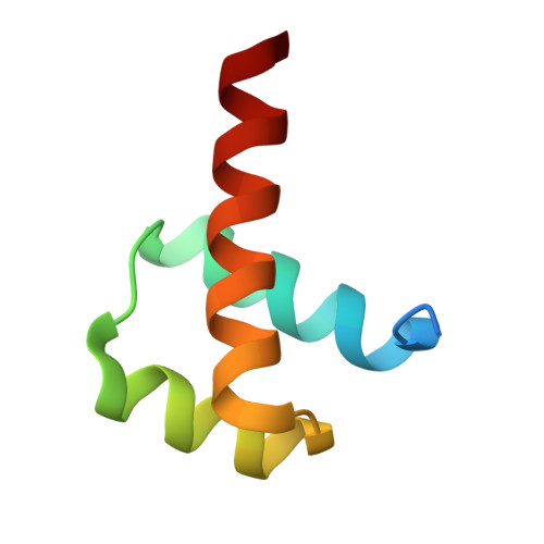 | |
UniProt & NIH Common Fund Data Resources | |||||
PHAROS: P43699 GTEx: ENSG00000136352 | |||||
Entity Groups | |||||
| Sequence Clusters | 30% Identity50% Identity70% Identity90% Identity95% Identity100% Identity | ||||
| UniProt Group | P43699 | ||||
Sequence AnnotationsExpand | |||||
Reference Sequence | |||||
Entity ID: 1 | ||||
| Molecule | Chains | Length | Organism | Image |
|---|---|---|---|---|
| CACG-containing 12bp forward strand | 12 | Homo sapiens | 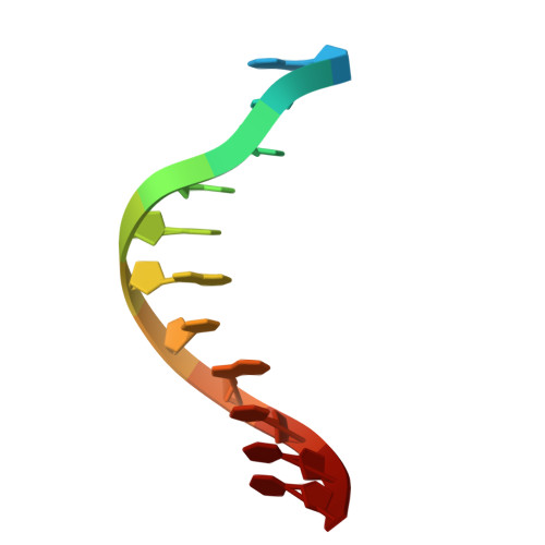 | |
Sequence AnnotationsExpand | ||||
Reference Sequence | ||||
Entity ID: 2 | ||||
| Molecule | Chains | Length | Organism | Image |
|---|---|---|---|---|
| 12bp reverse complementary | 12 | Homo sapiens | 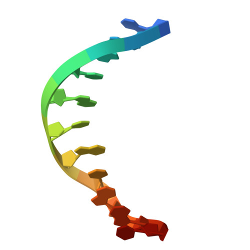 | |
Sequence AnnotationsExpand | ||||
Reference Sequence | ||||
| Ligands 1 Unique | |||||
|---|---|---|---|---|---|
| ID | Chains | Name / Formula / InChI Key | 2D Diagram | 3D Interactions | |
| MG Download:Ideal Coordinates CCD File | G [auth C] | MAGNESIUM ION Mg JLVVSXFLKOJNIY-UHFFFAOYSA-N |  | ||
| Length ( Å ) | Angle ( ˚ ) |
|---|---|
| a = 109.94 | α = 90 |
| b = 39.016 | β = 111.04 |
| c = 69.996 | γ = 90 |
| Software Name | Purpose |
|---|---|
| PHENIX | refinement |
| XDS | data scaling |
| PDB_EXTRACT | data extraction |
| XDS | data reduction |
| PHENIX | phasing |
| Funding Organization | Location | Grant Number |
|---|---|---|
| German Research Foundation (DFG) | Germany | FOR 2841 "Beyond the exome" |
| Polish National Science Centre | Poland | 3033/44/C/NZ1/00085 |


