Download Mendeley
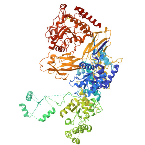
Structural basis of glycoprotein recognition by the human folding sensor UDP-glucose:glycoprotein glucosyltransferase.
Aparicio, D., Ortigosa, J.R., Guay, K.P., Williams, R.V., Lannot, J.O., Zong, G., Comino, N., Rodriguez, J.F., Wang, L.X., Marti, M.A., Roversi, P., Hebert, D.N., Gierasch, L.M., Modenutti, C.P., Guerin, M.E.(2026) Proc Natl Acad Sci U S A 123: e2621329123-e2621329123
- PubMed: 42961939 Search on PubMedSearch on PubMed Central
- DOI: https://doi.org/10.1073/pnas.2621329123
- Primary Citation Related Structures:
29DS, 9TV4 - PubMed Abstract:
UDP-glucose:glycoprotein glucosyltransferase 1 (UGGT1) plays a central role in quality control of glycoprotein folding within the early secretory pathway, yet the molecular basis of UGGT1 substrate recognition remains poorly understood. Here, we report cryo-EM structures of human full-length multidomain UGGT1. Monomeric UGGT1 structures reveal two conformational states corresponding to "open" and "closed" architectures. Three-dimensional variability and molecular dynamics simulations suggest the existence of intermediate conformations, leading to a model in which a continuum of structural transitions underlies UGGT1 function. Notably, in the open state, the EM-maps reveal at the top of the U-shaped protein a density that bridges the sensory TRXL2 and the glucosyltransferase catalytic domains, consistent with substrate association. This observation supports a mode of binding that places the substrate near the catalytic domain. A dimeric form of UGGT1 was observed in which the glycoprotein substrate binding sites are occluded, suggesting that dimerization may regulate UGGT1 activity. To better understand the spatial relationship between the site of misfolding and that of UGGT-mediated N-glycan reglucosylation, we identified N-glycosylation sites on endogenous glycoprotein substrates by glycoproteomics. No consensus sequence was found flanking modified sites, indicating that substrate recognition relies on mechanisms beyond primary sequence. Mutations in the UGGT1 gene cause a novel congenital disorder of glycosylation characterized by developmental delay and intellectual disability, and the human UGGT1 structure helps explain how disease-associated missense and deletion variants disrupt function. Together, our data provide insight into the relationship between UGGT1 structure and function, integrating enzyme conformational states with client glycoprotein interaction and processing.
- Structural Glycobiology Laboratory, Department of Structural and Molecular Biology, Institute of Molecular Biology of Barcelona, Spanish National Research Council, Barcelona Science Park, Barcelona, Catalonia 08028, Spain.
Organizational Affiliation: