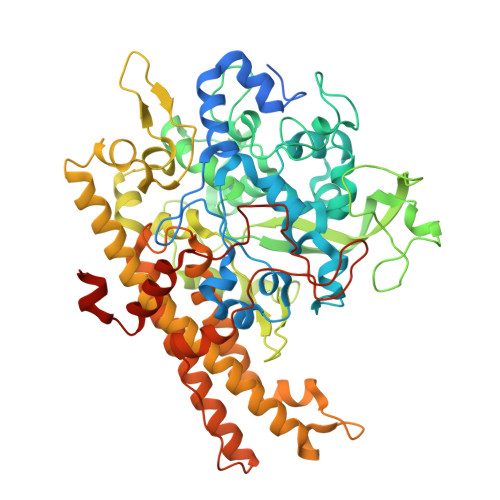
9TP1 | pdb_00009tp1

C. jejuni NrfA with Conventional His-Ligation at the Active-Site Heme


Experimental Data Snapshot

  • Method: X-RAY DIFFRACTION
  • Resolution: 1.60 Å
  • R-Value Free: 
    0.232 (Depositor), 0.232 (DCC) 
  • R-Value Work: 
    0.209 (Depositor), 0.208 (DCC) 
  • R-Value Observed: 
    0.210 (Depositor) 

wwPDB Validation 3D Report Full Report

Validation slider image for 9TP1

Ligand Structure Quality Assessment 


This is version 1.0 of the entry. See complete history. 

Literature

Lysine or histidine? examining the role of the proximal ligand at the active heme center of cytochrome c nitrite reductase.

Hermann, B., Rudolf, M., Messerschmidt, A., Simon, J., Einsle, O., Kroneck, P.M.H.

(2026) J Biol Inorg Chem 31: 317-328

  • DOI: https://doi.org/10.1007/s00775-026-02165-w
  • Primary Citation Related Structures: 
    9TP1, 9TP3, 9TP4

  • PubMed Abstract: 

    The identity of the proximal ligand of its catalytic heme 1 center - lysine or histidine - may directly affect how efficiently pentaheme cytochrome c nitrite reductase catalyzes the six-electron reduction of nitrite (NO 2 - ) to ammonium (NH 4 + ) and may also alter substrate specificity. This reaction is part of the pathway of dissimilatory nitrate reduction to ammonium, which competes with denitrification and avoids the formation of unreactive dinitrogen (N 2 ). In this study, we compare the biochemical properties and structures of cytochrome c nitrite reductase from Campylobacter jejuni with those of the enzyme from Wolinella succinogenes and examine its Lys134His variant. Notably, both the Campylobacter nitrite reductase and the Lys134His variant possess a catalytic heme 1 center coordinated by histidine as the proximal ligand, whereas the native Wolinella enzyme is coordinated by lysine. Despite this difference, both enzymes catalyze the reduction of NO 2 - to NH 4 + with high specific activities. To enable biochemical and structural characterization of these enzymes, we developed optimized protocols for protein expression and purification. The data we present allow, for the first time, a direct analysis of the influence of histidine or lysine as the proximal nitrogen ligand at the catalytic heme 1 center of pentaheme cytochrome c nitrite reductase within otherwise similar protein backgrounds.


  • Organizational Affiliation: 
    • Institute for Physical Chemistry, Albert-Ludwigs-University Freiburg, Albertstrasse 21, 79104, Freiburg, Germany.

Macromolecule Content 

  • Total Structure Weight: 147.92 kDa 
  • Atom Count: 10,532 
  • Modeled Residue Count: 1,123 
  • Deposited Residue Count: 1,240 
  • Unique protein chains: 1

Macromolecules

Find similar proteins by:|  3D Structure
Entity ID: 1
MoleculeChains  Sequence LengthOrganismDetailsImage
nitrite reductase (cytochrome; ammonia-forming)
A, B
620Campylobacter jejuniMutation(s): 0 
Gene Names: nrfA, Cj1357c
EC: 1.7.2.2
UniProt
Find proteins for Q0P8Q2 (Campylobacter jejuni subsp. jejuni serotype O:2 (strain ATCC 700819 / NCTC 11168))
Explore Q0P8Q2 
Go to UniProtKB:  Q0P8Q2
Entity Groups
Sequence Clusters30% Identity50% Identity70% Identity90% Identity95% Identity100% Identity
UniProt GroupQ0P8Q2
Sequence Annotations
Expand
Reference Sequence

Small Molecules

Ligands 5 Unique
IDChains Name / Formula / InChI Key2D Diagram3D Interactions
HEM
(Subject of Investigation/LOI)

Query on HEM


Download:Ideal Coordinates CCD File
F [auth A]
G [auth A]
H [auth A]
I [auth A]
J [auth A]
F [auth A],
G [auth A],
H [auth A],
I [auth A],
J [auth A],
N [auth B],
O [auth B],
P [auth B],
Q [auth B],
R [auth B]
PROTOPORPHYRIN IX CONTAINING FE
C34 H32 Fe N4 O4
KABFMIBPWCXCRK-RGGAHWMASA-L
P6G

Query on P6G


Download:Ideal Coordinates CCD File
S [auth B]HEXAETHYLENE GLYCOL
C12 H26 O7
IIRDTKBZINWQAW-UHFFFAOYSA-N
P4G

Query on P4G


Download:Ideal Coordinates CCD File
T [auth B]1-ETHOXY-2-(2-ETHOXYETHOXY)ETHANE
C8 H18 O3
RRQYJINTUHWNHW-UHFFFAOYSA-N
CA

Query on CA


Download:Ideal Coordinates CCD File
C [auth A]
D [auth A]
E [auth A]
K [auth B]
L [auth B]
C [auth A],
D [auth A],
E [auth A],
K [auth B],
L [auth B],
M [auth B]
CALCIUM ION
Ca
BHPQYMZQTOCNFJ-UHFFFAOYSA-N
NA

Query on NA


Download:Ideal Coordinates CCD File
U [auth B]SODIUM ION
Na
FKNQFGJONOIPTF-UHFFFAOYSA-N

Experimental Data & Validation

Experimental Data

  • Method: X-RAY DIFFRACTION
  • Resolution: 1.60 Å
  • R-Value Free:  0.232 (Depositor), 0.232 (DCC) 
  • R-Value Work:  0.209 (Depositor), 0.208 (DCC) 
  • R-Value Observed: 0.210 (Depositor) 
Space Group: P 1 21 1
Unit Cell:
Length ( Å )Angle ( ˚ )
a = 64.646α = 90
b = 158.965β = 117.17
c = 68γ = 90
Software Package:
Software NamePurpose
REFMACrefinement
Aimlessdata scaling
XDSdata reduction
MOLREPphasing
PDB_EXTRACTdata extraction

Structure Validation

View Full Validation Report


Ligand Structure Quality Assessment 


Entry History 

& Funding Information

Deposition Data


Funding OrganizationLocationGrant Number
German Research Foundation (DFG)Germany--

Revision History  (Full details and data files)

  • Version 1.0: 2026-10-28
    Type: Initial release