9TNZ | pdb_00009tnz

SP100 CARD filament


Experimental Data Snapshot

  • Method: ELECTRON MICROSCOPY
  • Resolution: 3.52 Å
  • Aggregation State: FILAMENT 
  • Reconstruction Method: HELICAL 

Starting Model: in silico
View more details

wwPDB Validation 3D Report Full Report

Validation slider image for 9TNZ

This is version 1.2 of the entry. See complete history

Literature

An SP110-SP100 axis is a critical regulator of promyelocytic leukaemia body dynamics and mitotic fidelity.

Aird, E.J.Rabl, J.Knuesel, T.Groen, K.Awwad, S.W.Korablev, B.Scherpe, L.Al-Herz, W.Hupfer, R.Recher, M.Jackson, S.P.Hale, B.G.Corn, J.E.

(2026) Nat Cell Biol 28: 684-695

  • DOI: https://doi.org/10.1038/s41556-026-01916-w
  • Primary Citation Related Structures: 
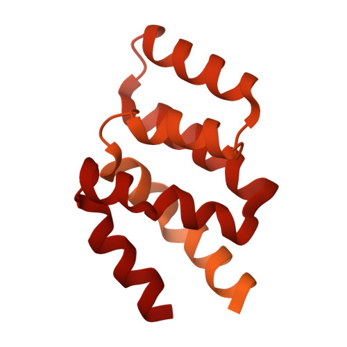
    9TNZ

  • PubMed Abstract: 

    Stimulation of the innate immune system by foreign RNA elicits a potent interferon response and can trigger cell death. The mechanisms by which cells balance a robust response with cell-intrinsic lethality are still being uncovered. Here, using genome-wide CRISPR-Cas9 genetic screens with triphosphorylated RNA stimulation, we discover that promyelocytic leukaemia (PML) nuclear body-localized speckled protein 110 (SP110) is a potent inhibitor of type 1 interferon-driven cell death. Death suppression by SP110 counteracts a toxic activity of SP100, a major constituent of PML bodies. Loss of SP110 leads to mitotic retention of SP100 and PML bodies, which associate with and perturb segregating chromosomes, leading to micronucleus formation, DNA damage and genotoxic cell death. A combination of cryo-electron microscopy, AlphaFold modelling and cellular biochemistry reveals that SP110 dissolves toxic SP100 oligomers via necessary and sufficient direct interactions between their caspase activation and recruitment domains. These data reveal the critical roles of SP100 and SP110 in governing the disassembly of PML bodies during mitosis, as well as the repercussions if this process is misregulated.


  • Organizational Affiliation
    • Institute of Molecular Health Sciences, Department of Biology, ETH Zurich, Zurich, Switzerland.

Macromolecule Content 

  • Total Structure Weight: 1,530.73 kDa 
  • Atom Count: 21,502 
  • Modeled Residue Count: 2,574 
  • Deposited Residue Count: 13,650 
  • Unique protein chains: 1

Macromolecules

Find similar proteins by:|  3D Structure
Entity ID: 1
MoleculeChains  Sequence LengthOrganismDetailsImage
Maltose/maltodextrin-binding periplasmic protein,Nuclear autoantigen Sp-100525Escherichia coli K-12Homo sapiens
This entity is chimeric
Mutation(s): 0 
Gene Names: malEb4034JW3994SP100
UniProt & NIH Common Fund Data Resources
Find proteins for P0AEX9 (Escherichia coli (strain K12))
Explore P0AEX9 
Go to UniProtKB:  P0AEX9
Find proteins for P23497 (Homo sapiens)
Explore P23497 
Go to UniProtKB:  P23497
PHAROS:  P23497
GTEx:  ENSG00000067066 
Entity Groups
Sequence Clusters30% Identity50% Identity70% Identity90% Identity95% Identity100% Identity
UniProt GroupsP23497P0AEX9
Sequence Annotations
Expand
Reference Sequence

Experimental Data & Validation

Experimental Data

  • Method: ELECTRON MICROSCOPY
  • Resolution: 3.52 Å
  • Aggregation State: FILAMENT 
  • Reconstruction Method: HELICAL 
EM Software:
TaskSoftware PackageVersion
MODEL REFINEMENTPHENIX1.21rc1_4985
RECONSTRUCTIONcryoSPARC4.4.0

Structure Validation

View Full Validation Report


Entry History 

& Funding Information

Deposition Data


Funding OrganizationLocationGrant Number
European Research Council (ERC)European Union855741-DDREAMM-ERC-2019-SyG
Swiss National Science FoundationSwitzerland310030_188858
European Molecular Biology Organization (EMBO)European UnionALTF 144-2021

Revision History  (Full details and data files)

  • Version 1.0: 2026-02-11
    Type: Initial release
  • Version 1.1: 2026-04-22
    Changes: Data collection, Database references
  • Version 1.2: 2026-04-29
    Changes: Data collection, Database references