9THY | pdb_00009thy

ReAIV structure containing an acrylamide adduct at Cys183 and an orthoborate ester at Ser47


Experimental Data Snapshot

  • Method: X-RAY DIFFRACTION
  • Resolution: 1.40 Å
  • R-Value Free: 
    0.183 (Depositor), 0.184 (DCC) 
  • R-Value Work: 
    0.137 (Depositor), 0.139 (DCC) 
  • R-Value Observed: 
    0.137 (Depositor) 

Starting Model: experimental
View more details

wwPDB Validation 3D Report Full Report

Validation slider image for 9THY

Ligand Structure Quality Assessment 


This is version 1.0 of the entry. See complete history

Literature

Efficient acrylamide adduct formation suggests dual applications of ReAIV L-asparaginase.

Sliwiak, J.Grzechowiak, M.Worsztynowicz, P.Pokrywka, K.Ruszkowski, M.Gilski, M.Jaskolski, M.

(2026) Amino Acids 

  • DOI: https://doi.org/10.1007/s00726-026-03525-1
  • Primary Citation Related Structures: 
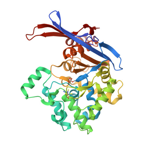
    9THY

  • PubMed Abstract: 

    Isothermal titration calorimetry (ITC) studies of the enzyme kinetics and substrate specificity of Rhizobium etli Class 3 L-asparaginases, ReAIV (constitutive) and ReAV (inducible), showed that despite highly conserved catalytic site, the two isoforms differ significantly in thermostability, zinc affinity, and biochemical properties. As part of a wider investigation of potential non-natural substrates, acrylamide was tested, revealing a pronounced heat effect with ReAIV but none with ReAV. Crystallographic analysis showed the formation of a Michael adduct between acrylamide and a surface-exposed cysteine 183 in ReAIV, while the catalytic activity toward L-asparagine hydrolysis remained unaffected. These findings highlight the unique and multimodal reactivity of ReAIV, suggesting its potential dual application in the food industry: in selective removal of L-asparagine and in covalent sequestration of acrylamide under mild conditions. The acrylamide modification improved crystal morphology of ReAIV, offering practical advantages for structural studies. Additionally, a covalent modification of the catalytic Ser47 residue was observed in the presented crystal structure. Based on B-factor analysis, literature data, and detection of borate contamination in the laboratory water, this modification was interpreted as an orthoborate ester.


  • Organizational Affiliation
    • Institute of Bioorganic Chemistry, Polish Academy of Sciences, Poznan, Poland. sliwiak@ibch.poznan.pl.

Macromolecule Content 

  • Total Structure Weight: 73.37 kDa 
  • Atom Count: 6,039 
  • Modeled Residue Count: 678 
  • Deposited Residue Count: 682 
  • Unique protein chains: 1

Macromolecules

Find similar proteins by:|  3D Structure
Entity ID: 1
MoleculeChains  Sequence LengthOrganismDetailsImage
L-asparaginase II protein
A, B
341Rhizobium etliMutation(s): 0 
Gene Names: RHE_CH01144
UniProt
Find proteins for Q2KB35 (Rhizobium etli (strain ATCC 51251 / DSM 11541 / JCM 21823 / NBRC 15573 / CFN 42))
Explore Q2KB35 
Go to UniProtKB:  Q2KB35
Entity Groups
Sequence Clusters30% Identity50% Identity70% Identity90% Identity95% Identity100% Identity
UniProt GroupQ2KB35
Sequence Annotations
Expand
Reference Sequence

Small Molecules

Ligands 5 Unique
IDChains Name / Formula / InChI Key2D Diagram3D Interactions
ROP
(Subject of Investigation/LOI)

Query on ROP


Download:Ideal Coordinates CCD File
P [auth A],
Y [auth B]
PROPIONAMIDE
C3 H7 N O
QLNJFJADRCOGBJ-UHFFFAOYSA-N
ZN

Query on ZN


Download:Ideal Coordinates CCD File
C [auth A],
Q [auth B]
ZINC ION
Zn
PTFCDOFLOPIGGS-UHFFFAOYSA-N
EDO

Query on EDO


Download:Ideal Coordinates CCD File
K [auth A],
L [auth A],
V [auth B],
W [auth B],
X [auth B]
1,2-ETHANEDIOL
C2 H6 O2
LYCAIKOWRPUZTN-UHFFFAOYSA-N
CL

Query on CL


Download:Ideal Coordinates CCD File
D [auth A]
E [auth A]
F [auth A]
G [auth A]
H [auth A]
D [auth A],
E [auth A],
F [auth A],
G [auth A],
H [auth A],
I [auth A],
J [auth A],
R [auth B],
S [auth B],
T [auth B],
U [auth B]
CHLORIDE ION
Cl
VEXZGXHMUGYJMC-UHFFFAOYSA-M
MG

Query on MG


Download:Ideal Coordinates CCD File
M [auth A],
N [auth A],
O [auth A]
MAGNESIUM ION
Mg
JLVVSXFLKOJNIY-UHFFFAOYSA-N
Modified Residues  1 Unique
IDChains TypeFormula2D DiagramParent
SEE
Query on SEE
A, B
L-PEPTIDE LINKINGC3 H9 B N O6SER

Experimental Data & Validation

Experimental Data

  • Method: X-RAY DIFFRACTION
  • Resolution: 1.40 Å
  • R-Value Free:  0.183 (Depositor), 0.184 (DCC) 
  • R-Value Work:  0.137 (Depositor), 0.139 (DCC) 
  • R-Value Observed: 0.137 (Depositor) 
Space Group: P 21 21 21
Unit Cell:
Length ( Å )Angle ( ˚ )
a = 61.415α = 90
b = 80.453β = 90
c = 142.715γ = 90
Software Package:
Software NamePurpose
PHENIXrefinement
PDB_EXTRACTdata extraction
XDSdata reduction
XDSdata scaling
PHASERphasing

Structure Validation

View Full Validation Report


Ligand Structure Quality Assessment 


Entry History 

& Funding Information

Deposition Data


Funding OrganizationLocationGrant Number
Polish National Science CentrePoland2020/37/B/NZ1/03250.

Revision History  (Full details and data files)

  • Version 1.0: 2026-05-27
    Type: Initial release