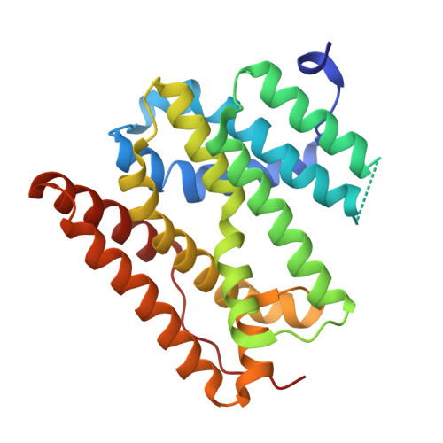
9TH3 | pdb_00009th3

Guanidine prenyltransferase NosH in complex with FSPP, guanidine and Mg2+


Experimental Data Snapshot

  • Method: X-RAY DIFFRACTION
  • Resolution: 2.10 Å
  • R-Value Free: 
    0.202 (Depositor), 0.202 (DCC) 
  • R-Value Work: 
    0.175 (Depositor), 0.179 (DCC) 
  • R-Value Observed: 
    0.177 (Depositor) 

Starting Model: in silico
View more details

wwPDB Validation 3D Report Full Report

Validation slider image for 9TH3

Ligand Structure Quality Assessment 


This is version 1.0 of the entry. See complete history. 

Literature

Natural product biosynthesis by guanidine prenyltransferases in plants and bacteria.

Kolling, D., Konzok, K., Taser, D., Jentsch, T., Schwoerer, S., Struwe, H., Bultemeier, A., Freiberg, J., Witte, C.P., Franke, J., Luzhetskyy, A., Kirschning, A., Rudorf, S., Koehnke, J.

(2026) Science 394: 413-418

  • DOI: https://doi.org/10.1126/science.aeg1821
  • Primary Citation Related Structures: 
    31KA, 9TGV, 9TGX, 9TGZ, 9TH3

  • PubMed Abstract: 

    Guanidine exists almost exclusively as a resonance-stabilized cation, making its direct enzymatic functionalization challenging. We identified the cyanobacterial enzyme NosH, which catalyzes direct prenyl transfer to free guanidine. Structural and mechanistic analyses revealed guanidine activation by a two-aspartate clamp coupled to prenyl-donor binding through a distinct catalytic architecture. Guided by these features, we identified a large family of guanidine prenyltransferases across bacteria and plants. Plant enzymes produced guanidine alkaloids such as galegine in vitro and heterologous expression of a plant ortholog in Nicotiana benthamiana produced galegine in planta, providing a direct enzymatic link to plant-specialized metabolism.


  • Organizational Affiliation: 
    • Institute of Food Chemistry, Leibniz University Hannover, Hannover, Germany.

Macromolecule Content 

  • Total Structure Weight: 60.47 kDa 
  • Atom Count: 4,248 
  • Modeled Residue Count: 475 
  • Deposited Residue Count: 516 
  • Unique protein chains: 1

Macromolecules

Find similar proteins by:|  3D Structure
Entity ID: 1
MoleculeChains  Sequence LengthOrganismDetailsImage
NosH
A, B
258Nostoc flagelliforme CCNUN1Mutation(s): 0 
Gene Names: COO91_01263
UniProt
Find proteins for A0A2K8SKL8 (Nostoc flagelliforme CCNUN1)
Explore A0A2K8SKL8 
Go to UniProtKB:  A0A2K8SKL8
Entity Groups
Sequence Clusters30% Identity50% Identity70% Identity90% Identity95% Identity100% Identity
UniProt GroupA0A2K8SKL8
Sequence Annotations
Expand
Reference Sequence

Small Molecules

Ligands 3 Unique
IDChains Name / Formula / InChI Key2D Diagram3D Interactions
FPS
(Subject of Investigation/LOI)

Query on FPS


Download:Ideal Coordinates CCD File
D [auth A],
H [auth B]
S-[(2E,6E)-3,7,11-TRIMETHYLDODECA-2,6,10-TRIENYL] TRIHYDROGEN THIODIPHOSPHATE
C15 H28 O6 P2 S
MYMLCRQRXFRQGP-YFVJMOTDSA-N
GAI
(Subject of Investigation/LOI)

Query on GAI


Download:Ideal Coordinates CCD File
C [auth A],
G [auth B]
GUANIDINE
C H5 N3
ZRALSGWEFCBTJO-UHFFFAOYSA-N
MG

Query on MG


Download:Ideal Coordinates CCD File
E [auth A],
F [auth A],
I [auth B],
J [auth B]
MAGNESIUM ION
Mg
JLVVSXFLKOJNIY-UHFFFAOYSA-N

Experimental Data & Validation

Experimental Data

  • Method: X-RAY DIFFRACTION
  • Resolution: 2.10 Å
  • R-Value Free:  0.202 (Depositor), 0.202 (DCC) 
  • R-Value Work:  0.175 (Depositor), 0.179 (DCC) 
  • R-Value Observed: 0.177 (Depositor) 
Space Group: I 1 2 1
Unit Cell:
Length ( Å )Angle ( ˚ )
a = 95.862α = 90
b = 77.383β = 106.022
c = 99.72γ = 90
Software Package:
Software NamePurpose
PHENIXrefinement
autoPROCdata reduction
Aimlessdata scaling
PHASERphasing

Structure Validation

View Full Validation Report


Ligand Structure Quality Assessment 


Entry History 

& Funding Information

Deposition Data


Funding OrganizationLocationGrant Number
Other governmentGermany--

Revision History  (Full details and data files)

  • Version 1.0: 2026-11-04
    Type: Initial release