9TGA | pdb_00009tga

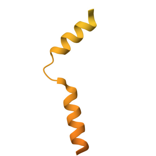
Drebrin actin binding domain 2 bound to F-actin


Experimental Data Snapshot

  • Method: ELECTRON MICROSCOPY
  • Resolution: 2.29 Å
  • Aggregation State: FILAMENT 
  • Reconstruction Method: SINGLE PARTICLE 

wwPDB Validation 3D Report Full Report

Validation slider image for 9TGA

This is version 1.0 of the entry. See complete history

Literature

Structural mechanisms of drebrin-mediated F-actin network modulation.

Zhao, W.Chu, L.Y.Abis, G.Oozeer, F.Mulvaney, T.Nagar, N.Topf, M.Gordon-Weeks, P.R.Conte, M.R.Atherton, J.

(2026) Nat Commun 17

  • DOI: https://doi.org/10.1038/s41467-026-74543-6
  • Primary Citation Related Structures: 
    9TG8, 9TG9, 9TGA

  • PubMed Abstract: 

    Drebrin modulates F-actin networks and links them to other intracellular components, regulating crucial processes including neuritogenesis, synaptic plasticity, virus internalisation and cancer invasion. Using single-particle cryo-EM we characterise drebrin's interaction with F-actin through two separate conserved actin binding domains (ABD1 and ABD2), revealing structural bases for its F-actin-modulating properties. We describe a multimodal interaction where drebrin's ABD1 can adopt two conformations and a long flexible loop connecting to ABD2 allows the two ABDs to occupy multiple relative positions along F-actin. The flexible loop connecting the two ABDs also confers some propensity to loosely bundle F-actin. Drebrin's ABDs bind across multiple actin protomers and their subdomains and modify the longitudinal inter-protomer interface, explaining its F-actin stabilising properties. Furthermore, we show drebrin's binding site on F-actin is shared with other critical actin-binding and regulatory proteins, explaining their competitive displacement.


  • Organizational Affiliation
    • Randall Centre for Cell and Molecular Biophysics, King's College London - New Hunt's House, Guy's Campus, London, UK.

Macromolecule Content 

  • Total Structure Weight: 111.31 kDa 
  • Atom Count: 6,432 
  • Modeled Residue Count: 770 
  • Deposited Residue Count: 975 
  • Unique protein chains: 2

Macromolecules

Find similar proteins by:|  3D Structure
Entity ID: 1
MoleculeChains  Sequence LengthOrganismDetailsImage
Drebrin221Homo sapiensMutation(s): 0 
Gene Names: DBN1D0S117E
UniProt & NIH Common Fund Data Resources
Find proteins for Q16643 (Homo sapiens)
Explore Q16643 
Go to UniProtKB:  Q16643
PHAROS:  Q16643
GTEx:  ENSG00000113758 
Entity Groups
Sequence Clusters30% Identity50% Identity70% Identity90% Identity95% Identity100% Identity
UniProt GroupQ16643
Sequence Annotations
Expand
Reference Sequence
Find similar proteins by:|  3D Structure
Entity ID: 2
MoleculeChains  Sequence LengthOrganismDetailsImage
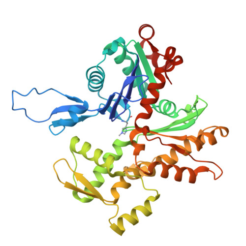
Actin, alpha skeletal muscleB,
C [auth D]
377Oryctolagus cuniculusMutation(s): 0 
EC: 3.6.4
UniProt
Find proteins for P68135 (Oryctolagus cuniculus)
Explore P68135 
Go to UniProtKB:  P68135
Entity Groups
Sequence Clusters30% Identity50% Identity70% Identity90% Identity95% Identity100% Identity
UniProt GroupP68135
Sequence Annotations
Expand
Reference Sequence

Experimental Data & Validation

Experimental Data

  • Method: ELECTRON MICROSCOPY
  • Resolution: 2.29 Å
  • Aggregation State: FILAMENT 
  • Reconstruction Method: SINGLE PARTICLE 
EM Software:
TaskSoftware PackageVersion
RECONSTRUCTIONRELION5.0
MODEL REFINEMENTPHENIX1.21.2_5419

Structure Validation

View Full Validation Report


Entry History 

& Funding Information

Deposition Data


Funding OrganizationLocationGrant Number
Biotechnology and Biological Sciences Research Council (BBSRC)United KingdomBB/V006568/1
UK Research and Innovation (UKRI)United KingdomUKRI2979
Leverhulme TrustUnited KingdomRPG-2020264
Wellcome TrustUnited Kingdom209250/Z/17/ Z
Wellcome TrustUnited Kingdom206175/Z/17/Z
Wellcome TrustUnited Kingdom202767/Z/16/Z
Leverhulme TrustUnited KingdomEM-2022-038-2
British Heart FoundationUnited KingdomIG/16/2/32273

Revision History  (Full details and data files)

  • Version 1.0: 2026-09-02
    Type: Initial release