9TBJ | pdb_00009tbj

Leishmania mexicana secreted acid phosphatase


Experimental Data Snapshot

  • Method: ELECTRON MICROSCOPY
  • Resolution: 3.00 Å
  • Aggregation State: FILAMENT 
  • Reconstruction Method: SINGLE PARTICLE 

Starting Model: in silico
View more details

wwPDB Validation 3D Report Full Report

Validation slider image for 9TBJ

This is version 1.1 of the entry. See complete history

Literature

Structural basis of secreted acid phosphatase polymerization in the Leishmania parasite.

Bose, P.Dahan, I.Upcher, A.Zalk, R.Grossman-Haham, I.

(2026) Protein Sci 35: e70591-e70591

  • DOI: https://doi.org/10.1002/pro.70591
  • Primary Citation Related Structures: 
    28IK, 9TBJ

  • PubMed Abstract: 

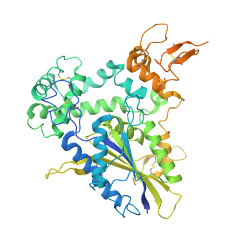
    Enzymes that assemble into filaments typically transition between protomeric and polymeric states in response to cellular conditions. In contrast, the secreted acid phosphatase (SAP) of Leishmania, one of the most abundant extracellular glycoproteins produced by the parasite and regarded as a major virulence factor in the neglected tropical disease leishmaniasis, exhibits fundamentally different behavior. Depending on the species, SAP forms either highly stable extracellular filaments or remains exclusively as globular particles, with no evidence of reversible interconversion. This binary assembly pattern is particularly intriguing given that SAP orthologs that differ in their ability to polymerize share a high degree of sequence conservation, leaving the molecular determinants of filament formation unknown. Here, we report the cryo-EM structure of filamentous Leishmania mexicana SAP to a global resolution of 3.0 Å. The structure resolves the multilevel organization of the enzyme, from individual catalytic phosphatase domains and their unique substrate-binding pockets to the formation of homodimeric protomers, the decoration with N-linked glycans, and the supramolecular organization into filaments. At the core of the polymerization interface, we identified a unique β-hairpin motif that has not been observed in any other phosphatase or enzyme filament, which provides exceptional filament stability. By integrating structural data with comparative sequence analysis and machine-learning-based structure predictions, we define the molecular basis for the species-specific assembly behaviors observed across Leishmania SAPs. This work establishes the principles governing SAP filament formation and provides a framework for understanding its evolution, enzymatic function, and potential applications.


  • Organizational Affiliation
    • Department of Life Sciences, Ben-Gurion University of the Negev, Beer Sheva, Israel.

Macromolecule Content 

  • Total Structure Weight: 192.28 kDa 
  • Atom Count: 10,259 
  • Modeled Residue Count: 1,267 
  • Deposited Residue Count: 1,695 
  • Unique protein chains: 1

Macromolecules

Find similar proteins by:|  3D Structure
Entity ID: 1
MoleculeChains  Sequence LengthOrganismDetailsImage
Secreted acid phosphatase 1 (SAP1)
A, B, C
565Leishmania mexicanaMutation(s): 0 
Gene Names: lmsap1
UniProt
Find proteins for Q25332 (Leishmania mexicana)
Explore Q25332 
Go to UniProtKB:  Q25332
Entity Groups
Sequence Clusters30% Identity50% Identity70% Identity90% Identity95% Identity100% Identity
UniProt GroupQ25332
Glycosylation
Glycosylation Sites: 5
Sequence Annotations
Expand
Reference Sequence

Oligosaccharides

Help  
Entity ID: 2
MoleculeChains Length2D Diagram GlycosylationD Interactions
alpha-D-mannopyranose-(1-4)-2-acetamido-2-deoxy-beta-D-glucopyranose-(1-4)-2-acetamido-2-deoxy-beta-D-glucopyranoseD,
G [auth I]
3N-Glycosylation
Glycosylation Resources
GlyTouCan: G62182OO
GlyCosmos: G62182OO
GlyGen: G62182OO
Entity ID: 3
MoleculeChains Length2D Diagram GlycosylationD Interactions
2-acetamido-2-deoxy-beta-D-glucopyranose-(1-4)-2-acetamido-2-deoxy-beta-D-glucopyranose
E [auth F],
F [auth G],
H [auth K],
I [auth L],
J [auth M],
E [auth F],
F [auth G],
H [auth K],
I [auth L],
J [auth M],
K [auth P],
L [auth Q]
2N-Glycosylation
Glycosylation Resources
GlyTouCan: G42666HT
GlyCosmos: G42666HT
GlyGen: G42666HT

Small Molecules

Ligands 2 Unique
IDChains Name / Formula / InChI Key2D Diagram3D Interactions
NAG
(Subject of Investigation/LOI)

Query on NAG


Download:Ideal Coordinates CCD File
M [auth A]
N [auth A]
P [auth B]
R [auth C]
S [auth C]
M [auth A],
N [auth A],
P [auth B],
R [auth C],
S [auth C],
T [auth C]
2-acetamido-2-deoxy-beta-D-glucopyranose
C8 H15 N O6
OVRNDRQMDRJTHS-FMDGEEDCSA-N
PO4
(Subject of Investigation/LOI)

Query on PO4


Download:Ideal Coordinates CCD File
O [auth A],
Q [auth B],
U [auth C]
PHOSPHATE ION
O4 P
NBIIXXVUZAFLBC-UHFFFAOYSA-K

Experimental Data & Validation

Experimental Data

  • Method: ELECTRON MICROSCOPY
  • Resolution: 3.00 Å
  • Aggregation State: FILAMENT 
  • Reconstruction Method: SINGLE PARTICLE 
EM Software:
TaskSoftware PackageVersion
MODEL REFINEMENTPHENIX1.21rc1_4985

Structure Validation

View Full Validation Report


Entry History 

& Funding Information

Deposition Data


Funding OrganizationLocationGrant Number
Israel Science FoundationIsrael1691/23

Revision History  (Full details and data files)

  • Version 1.0: 2026-04-29
    Type: Initial release
  • Version 1.1: 2026-05-06
    Changes: Data collection, Database references