9TBF | pdb_00009tbf

Cryo-EM structure of the light-driven sodium pump ErNaR in the monomeric form in the O2 state


Experimental Data Snapshot

  • Method: ELECTRON MICROSCOPY
  • Resolution: 3.30 Å
  • Aggregation State: PARTICLE 
  • Reconstruction Method: SINGLE PARTICLE 

wwPDB Validation 3D Report Full Report

Validation slider image for 9TBF

This is version 1.0 of the entry. See complete history

Literature

Microsecond Time-Resolved Cryo-EM Based on Jet Vitrification.

Haubner, M.Williams, H.M.Hruby, J.Straub, M.S.Guskov, A.Kovalev, K.Drabbels, M.Lorenz, U.J.

(2025) bioRxiv 

  • DOI: https://doi.org/10.1101/2025.11.21.689681
  • Primary Citation Related Structures: 
    9TBD, 9TBE, 9TBF

  • PubMed Abstract: 

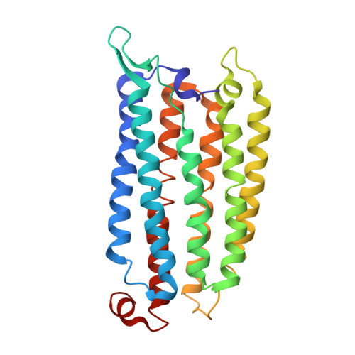
    Understanding and ultimately predicting the function of proteins is one of the frontiers in structural biology. This will only be possible if it becomes feasible to routinely observe proteins on the fast timescales on which they perform their tasks. Recently, laser flash melting and revitrification experiments have improved the time resolution of cryo-electron microscopy (cryo-EM) to microseconds, rendering it fast enough to observe the domain motions of proteins that are frequently linked to function. However, observations have been limited to a time window of just a few hundred microseconds. Here, we introduce time-resolved cryo-EM experiments based on jet vitrification that combine microsecond resolution with an observation window of up to seconds. We use a short laser pulse to initiate protein dynamics, and as they unfold, vitrify the sample with a jet of a liquid cryogen to arrest the dynamics at that point in time. We demonstrate that our approach affords near-atomic spatial resolution and a time resolution of 21 µs. This allows us to observe the photoinduced dynamics of the light-driven sodium pump Er NaR on the microsecond to millisecond timescale. Our experiments significantly expand the ability of cryo-EM to observe protein dynamics across multiple timescales.

Macromolecule Content 

  • Total Structure Weight: 35.32 kDa 
  • Atom Count: 2,409 
  • Modeled Residue Count: 274 
  • Deposited Residue Count: 274 
  • Unique protein chains: 1

Macromolecules

Find similar proteins by:|  3D Structure
Entity ID: 1
MoleculeChains  Sequence LengthOrganismDetailsImage
Bacteriorhodopsin-like protein274ErythrobacterMutation(s): 0 

Small Molecules

Ligands 3 Unique
IDChains Name / Formula / InChI Key2D Diagram3D Interactions
LMT

Query on LMT


Download:Ideal Coordinates CCD File
L [auth A],
M [auth A],
N [auth A]
DODECYL-BETA-D-MALTOSIDE
C24 H46 O11
NLEBIOOXCVAHBD-QKMCSOCLSA-N
RET
(Subject of Investigation/LOI)

Query on RET


Download:Ideal Coordinates CCD File
O [auth A]RETINAL
C20 H28 O
NCYCYZXNIZJOKI-OVSJKPMPSA-N
LFA

Query on LFA


Download:Ideal Coordinates CCD File
B [auth A]
C [auth A]
D [auth A]
E [auth A]
F [auth A]
B [auth A],
C [auth A],
D [auth A],
E [auth A],
F [auth A],
G [auth A],
H [auth A],
I [auth A],
J [auth A],
K [auth A]
EICOSANE
C20 H42
CBFCDTFDPHXCNY-UHFFFAOYSA-N

Experimental Data & Validation

Experimental Data

  • Method: ELECTRON MICROSCOPY
  • Resolution: 3.30 Å
  • Aggregation State: PARTICLE 
  • Reconstruction Method: SINGLE PARTICLE 
EM Software:
TaskSoftware PackageVersion
MODEL REFINEMENTPHENIX1.21.2_5419
RECONSTRUCTIONcryoSPARC4.6.0

Structure Validation

View Full Validation Report


Entry History 

& Funding Information

Deposition Data


Funding OrganizationLocationGrant Number
Swiss National Science FoundationSwitzerlandTMCG-2+213773

Revision History  (Full details and data files)

  • Version 1.0: 2025-12-17
    Type: Initial release