9TAN | pdb_00009tan

Local refinement of E. coli Complex I D79N NuoA mutant hydrophilic domain in LMNG


Experimental Data Snapshot

  • Method: ELECTRON MICROSCOPY
  • Resolution: 2.59 Å
  • Aggregation State: PARTICLE 
  • Reconstruction Method: SINGLE PARTICLE 

Starting Model: experimental
View more details

wwPDB Validation 3D Report Full Report

Validation slider image for 9TAN

This is version 1.1 of the entry. See complete history

Literature

A carboxylate switch point controls long-range energy transduction in respiratory Complex I.

Beghiah, A.Saura, P.Kovalova, T.Hoeser, F.Friedrich, T.Kaila, V.R.I.

(2026) Nat Commun 17

  • DOI: https://doi.org/10.1038/s41467-026-74767-6
  • Primary Citation Related Structures: 
    9TAJ, 9TAK, 9TAL, 9TAM, 9TAN, 9TAO

  • PubMed Abstract: 

    Complex I is a highly intricate membrane-bound protein complex that powers the cellular energy metabolism by a long-range ( > 300 Å) proton-coupled electron transfer (PCET) reaction. Here, we investigate the highly debated coupling mechanism of Complex I by probing the charge transfer reaction along its functionally central carboxylate pathway (E-channel). By combining biophysical and site-directed mutagenesis experiments with high-resolution (2.6-2.8 Å) cryo-electron microscopy (cryo-EM) and multiscale simulations, we identify a conserved carboxylate switch point (D79 NuoA ) that mediates proton transfer by establishing a kinetic gate and couples the redox chemistry to proton pumping. We find that mutation of the identified site, as found in patients suffering from severe neurodegenerative disorders, drastically perturbs the charge transfer mechanism, and results in a 20% PCET activity. Our combined findings illustrate mechanistic principles of molecular gates underlying long-range charge transfer reactions, and show how disease mutations perturb the function of conserved switch points in energy transduction.


  • Organizational Affiliation
    • Department of Biochemistry and Biophysics, Stockholm University, Stockholm, Sweden.

Macromolecule Content 

  • Total Structure Weight: 288.34 kDa 
  • Atom Count: 19,678 
  • Modeled Residue Count: 2,449 
  • Deposited Residue Count: 2,537 
  • Unique protein chains: 6

Macromolecules

Find similar proteins by:|  3D Structure
Entity ID: 1
MoleculeChains  Sequence LengthOrganismDetailsImage
NADH-quinone oxidoreductase subunit BA [auth B]220Escherichia coli BW25113Mutation(s): 0 
Gene Names: nuoBb2287JW5875
EC: 7.1.1
UniProt
Find proteins for P0AFC7 (Escherichia coli (strain K12))
Explore P0AFC7 
Go to UniProtKB:  P0AFC7
Entity Groups
Sequence Clusters30% Identity50% Identity70% Identity90% Identity95% Identity100% Identity
UniProt GroupP0AFC7
Sequence Annotations
Expand
Reference Sequence
Find similar proteins by:|  3D Structure
Entity ID: 2
MoleculeChains  Sequence LengthOrganismDetailsImage
NADH-quinone oxidoreductase subunit C/DB [auth C]600Escherichia coli BW25113Mutation(s): 0 
Gene Names: nuoCnuoCDnuoDb2286JW5375
EC: 7.1.1
UniProt
Find proteins for P33599 (Escherichia coli (strain K12))
Explore P33599 
Go to UniProtKB:  P33599
Entity Groups
Sequence Clusters30% Identity50% Identity70% Identity90% Identity95% Identity100% Identity
UniProt GroupP33599
Sequence Annotations
Expand
Reference Sequence
Find similar proteins by:|  3D Structure
Entity ID: 3
MoleculeChains  Sequence LengthOrganismDetailsImage
NADH-quinone oxidoreductase subunit EC [auth E]166Escherichia coli BW25113Mutation(s): 0 
Gene Names: nuoEb2285JW2280
EC: 7.1.1
UniProt
Find proteins for P0AFD1 (Escherichia coli (strain K12))
Explore P0AFD1 
Go to UniProtKB:  P0AFD1
Entity Groups
Sequence Clusters30% Identity50% Identity70% Identity90% Identity95% Identity100% Identity
UniProt GroupP0AFD1
Sequence Annotations
Expand
Reference Sequence
Find similar proteins by:|  3D Structure
Entity ID: 4
MoleculeChains  Sequence LengthOrganismDetailsImage
NADH-quinone oxidoreductase subunit FD [auth F]461Escherichia coli BW25113Mutation(s): 0 
Gene Names: nuoFb2284JW2279
EC: 7.1.1
UniProt
Find proteins for P31979 (Escherichia coli (strain K12))
Explore P31979 
Go to UniProtKB:  P31979
Entity Groups
Sequence Clusters30% Identity50% Identity70% Identity90% Identity95% Identity100% Identity
UniProt GroupP31979
Sequence Annotations
Expand
Reference Sequence
Find similar proteins by:|  3D Structure
Entity ID: 5
MoleculeChains  Sequence LengthOrganismDetailsImage
NADH-quinone oxidoreductase subunit GE [auth G]910Escherichia coli BW25113Mutation(s): 0 
Gene Names: nuoGb2283JW2278
EC: 7.1.1
UniProt
Find proteins for P33602 (Escherichia coli (strain K12))
Explore P33602 
Go to UniProtKB:  P33602
Entity Groups
Sequence Clusters30% Identity50% Identity70% Identity90% Identity95% Identity100% Identity
UniProt GroupP33602
Sequence Annotations
Expand
Reference Sequence
Find similar proteins by:|  3D Structure
Entity ID: 6
MoleculeChains  Sequence LengthOrganismDetailsImage
NADH-quinone oxidoreductase subunit IF [auth I]180Escherichia coli BW25113Mutation(s): 0 
Gene Names: nuoIb2281JW2276
EC: 7.1.1
UniProt
Find proteins for P0AFD6 (Escherichia coli (strain K12))
Explore P0AFD6 
Go to UniProtKB:  P0AFD6
Entity Groups
Sequence Clusters30% Identity50% Identity70% Identity90% Identity95% Identity100% Identity
UniProt GroupP0AFD6
Sequence Annotations
Expand
Reference Sequence

Small Molecules

Ligands 4 Unique
IDChains Name / Formula / InChI Key2D Diagram3D Interactions
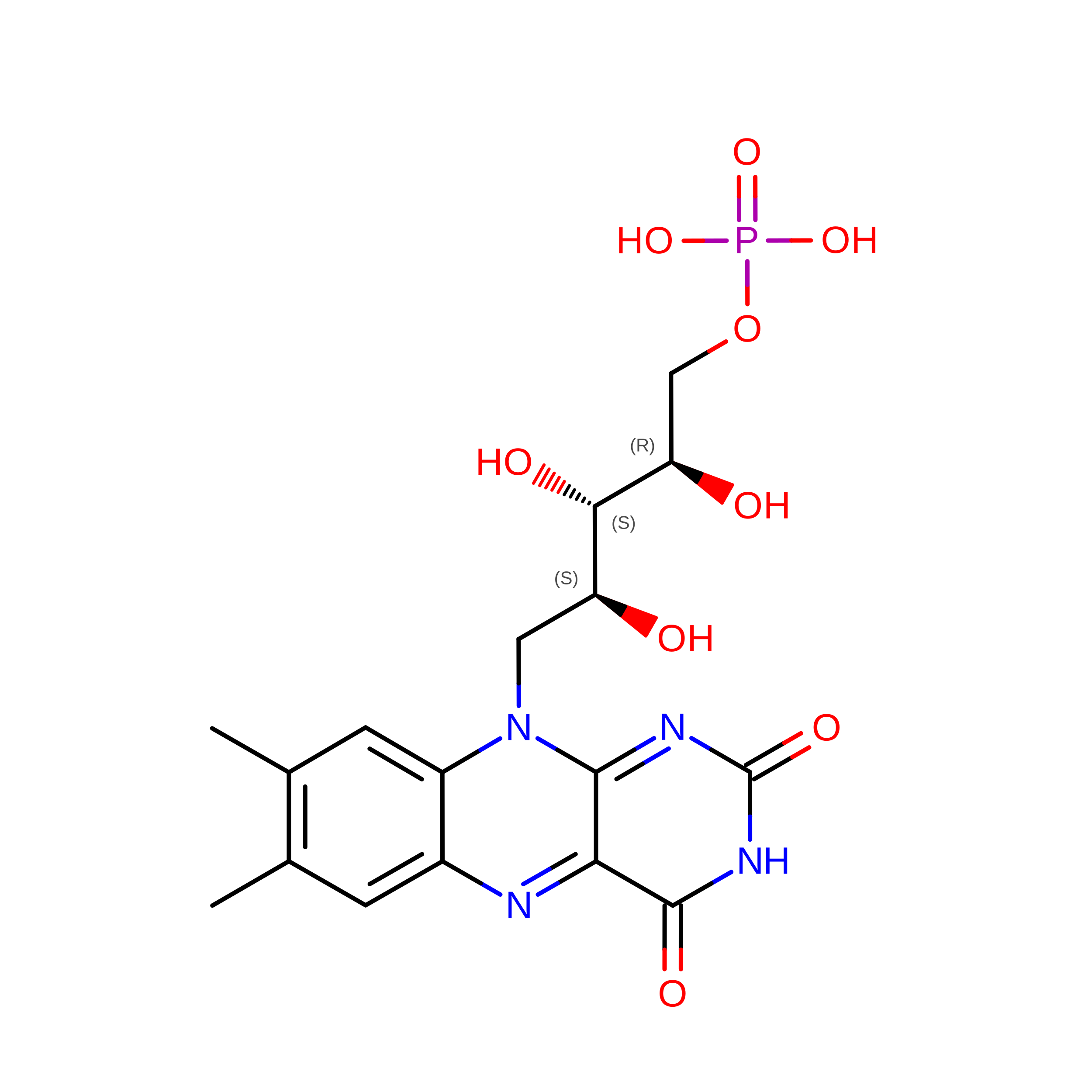
FMN

Query on FMN


Download:Ideal Coordinates CCD File
K [auth F]FLAVIN MONONUCLEOTIDE
C17 H21 N4 O9 P
FVTCRASFADXXNN-SCRDCRAPSA-N
SF4
(Subject of Investigation/LOI)

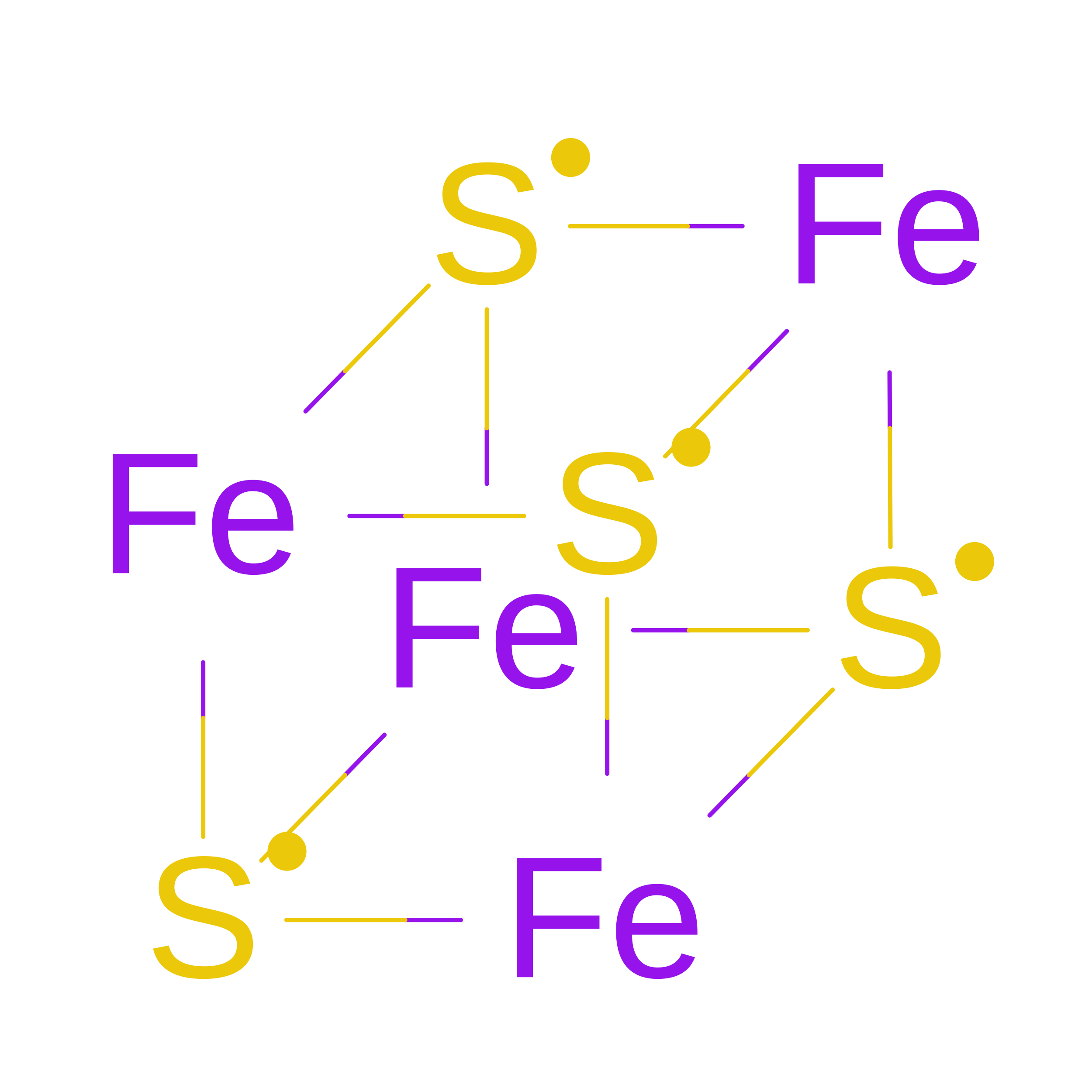
Query on SF4


Download:Ideal Coordinates CCD File
G [auth B]
J [auth F]
L [auth G]
M [auth G]
N [auth G]
G [auth B],
J [auth F],
L [auth G],
M [auth G],
N [auth G],
R [auth I],
S [auth I]
IRON/SULFUR CLUSTER
Fe4 S4
LJBDFODJNLIPKO-UHFFFAOYSA-N
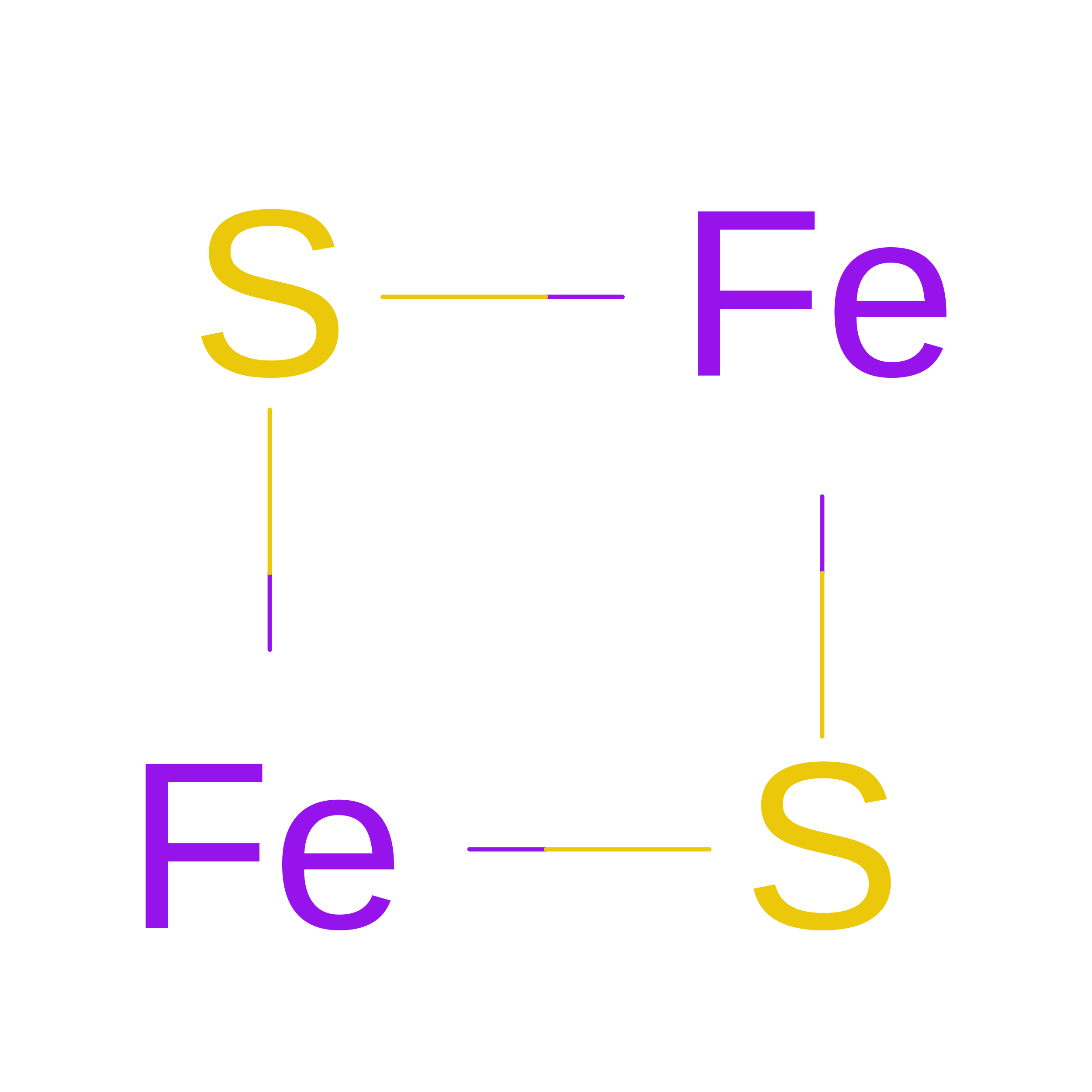
FES
(Subject of Investigation/LOI)

Query on FES


Download:Ideal Coordinates CCD File
H [auth E],
O [auth G]
FE2/S2 (INORGANIC) CLUSTER
Fe2 S2
NIXDOXVAJZFRNF-UHFFFAOYSA-N
CA

Query on CA


Download:Ideal Coordinates CCD File
I [auth E],
P [auth G],
Q [auth G],
T [auth I]
CALCIUM ION
Ca
BHPQYMZQTOCNFJ-UHFFFAOYSA-N

Experimental Data & Validation

Experimental Data

  • Method: ELECTRON MICROSCOPY
  • Resolution: 2.59 Å
  • Aggregation State: PARTICLE 
  • Reconstruction Method: SINGLE PARTICLE 
EM Software:
TaskSoftware PackageVersion
MODEL REFINEMENTPHENIX1.21.2_5419
RECONSTRUCTIONcryoSPARC

Structure Validation

View Full Validation Report


Entry History 

& Funding Information

Deposition Data


Funding OrganizationLocationGrant Number
Knut and Alice Wallenberg FoundationSweden2024.0220
Swedish Research CouncilSweden2020-04081

Revision History  (Full details and data files)

  • Version 1.0: 2026-05-06
    Type: Initial release
  • Version 1.1: 2026-07-15
    Changes: Data collection, Database references