9SQN | pdb_00009sqn

Structure of a disulfide-bridged complex between HLA-A*02:01-K127N/Y84C and ERp57/tapasin-K16C - State 1


Experimental Data Snapshot

  • Method: ELECTRON MICROSCOPY
  • Resolution: 3.50 Å
  • Aggregation State: PARTICLE 
  • Reconstruction Method: SINGLE PARTICLE 

wwPDB Validation 3D Report Full Report

Validation slider image for 9SQN

This is version 1.1 of the entry. See complete history. 

Literature

Dynamic states of an MHC class I molecule during peptide exchange.

Mitlehner, A., Pasos-Trejo, A.S., Becker, M., Lan, H., Berg, L., Hilal, T., Loll, B., Kuropka, B., Clementi, C., Freund, C.

(2026) Nat Commun 17

  • DOI: https://doi.org/10.1038/s41467-026-77731-6
  • Primary Citation Related Structures: 
    9SQM, 9SQN, 9SQO, 9SQP

  • PubMed Abstract: 

    Antigens of intracellular origin are processed then presented by proteins of the highly polymorphic major histocompatibility complex I (MHCI), thereby enabling T cell activation during an immune response. In particular, the peptide-exchange catalyst tapasin (Tsn) plays a critical role in shaping the pool of peptide antigens that ultimately reach the cell surface. Here, using disulfide bond engineering in conjunction with cryogenic electron microscopy (cryoEM) and molecular dynamics simulations, we provide evidence for partial collapse of the antigen binding groove during the peptide exchange process. An intermediate is formed that is characterized by interactions between the peptide's N-terminus and conserved tyrosine side chains in MHCI. Unfolding of the MHCI α1-helix in the Tsn-bound state is contrasted by a stable α2-helix that is kept in its native-like major conformation by the support of the large interface it entertains with the exchange catalyst. Helical disorder propensities and backbone flexibilities of the α1-helix are predicted to have increased during evolution, suggesting that the dynamic features introduced by polymorphic variation may have contributed to shaping the pool of antigens presented to T cells.


  • Organizational Affiliation: 
    • Laboratory of Protein Biochemistry, Institute of Chemistry and Biochemistry, Freie Universität Berlin, Berlin, Germany.

Macromolecule Content 

  • Total Structure Weight: 144.83 kDa 
  • Atom Count: 9,785 
  • Modeled Residue Count: 1,231 
  • Deposited Residue Count: 1,287 
  • Unique protein chains: 5

Macromolecules

Find similar proteins by:|  3D Structure
Entity ID: 1
MoleculeChains  Sequence LengthOrganismDetailsImage
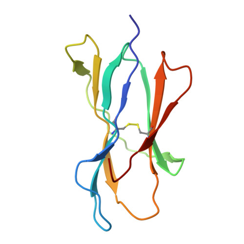
Beta-2-microglobulinA [auth B]99Homo sapiensMutation(s): 1 
Gene Names: B2M, CDABP0092, HDCMA22P
UniProt & NIH Common Fund Data Resources
Find proteins for P61769 (Homo sapiens)
Explore P61769 
Go to UniProtKB:  P61769
PHAROS:  P61769
GTEx:  ENSG00000166710 
Entity Groups
Sequence Clusters30% Identity50% Identity70% Identity90% Identity95% Identity100% Identity
UniProt GroupP61769
Sequence Annotations
Expand
Reference Sequence
Find similar proteins by:|  3D Structure
Entity ID: 2
MoleculeChains  Sequence LengthOrganismDetailsImage
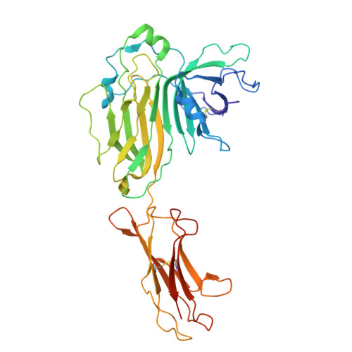
TapasinB [auth D]398Homo sapiensMutation(s): 3 
Gene Names: TAPBP, NGS17, TAPA
UniProt & NIH Common Fund Data Resources
Find proteins for O15533 (Homo sapiens)
Explore O15533 
Go to UniProtKB:  O15533
PHAROS:  O15533
GTEx:  ENSG00000231925 
Entity Groups
Sequence Clusters30% Identity50% Identity70% Identity90% Identity95% Identity100% Identity
UniProt GroupO15533
Glycosylation
Glycosylation Sites: 1Go to GlyGen: O15533-1
Sequence Annotations
Expand
Reference Sequence
Find similar proteins by:|  3D Structure
Entity ID: 3
MoleculeChains  Sequence LengthOrganismDetailsImage
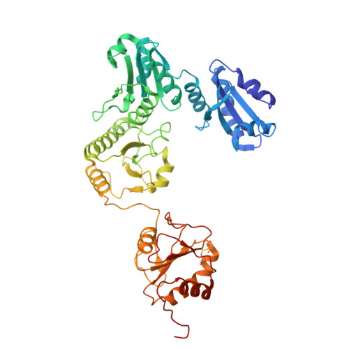
Protein disulfide-isomerase A3C [auth E]481Homo sapiensMutation(s): 1 
Gene Names: PDIA3, ERP57, ERP60, GRP58
EC: 5.3.4.1
UniProt & NIH Common Fund Data Resources
Find proteins for P30101 (Homo sapiens)
Explore P30101 
Go to UniProtKB:  P30101
PHAROS:  P30101
GTEx:  ENSG00000167004 
Entity Groups
Sequence Clusters30% Identity50% Identity70% Identity90% Identity95% Identity100% Identity
UniProt GroupP30101
Sequence Annotations
Expand
Reference Sequence
Find similar proteins by:|  3D Structure
Entity ID: 4
MoleculeChains  Sequence LengthOrganismDetailsImage
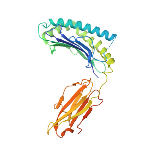
MHC class I antigenD [auth A]301Homo sapiensMutation(s): 1 
Gene Names: HLA-A
UniProt
Find proteins for Q9TQB6 (Homo sapiens)
Explore Q9TQB6 
Go to UniProtKB:  Q9TQB6
Entity Groups
Sequence Clusters30% Identity50% Identity70% Identity90% Identity95% Identity100% Identity
UniProt GroupQ9TQB6
Sequence Annotations
Expand
Reference Sequence
Find similar proteins by:  Sequence   |   3D Structure  
Entity ID: 5
MoleculeChains  Sequence LengthOrganismDetailsImage
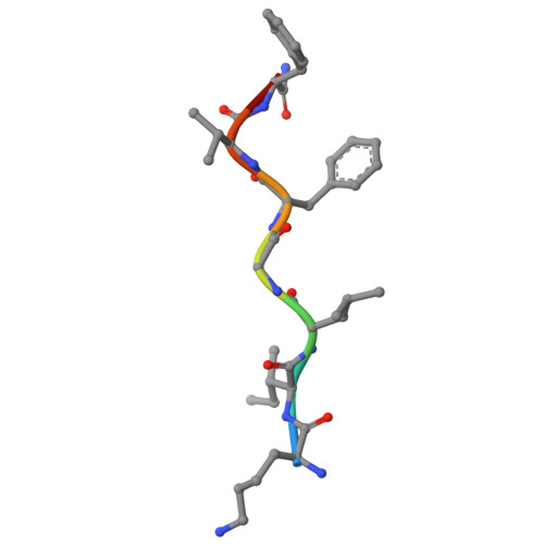
LYS-ILE-LEU-GLY-PHE-VAL-NFA, pKV9E [auth C]8Homo sapiensMutation(s): 0 
Sequence Annotations
Expand
Reference Sequence

Oligosaccharides

Help  
Entity ID: 6
MoleculeChains Length2D Diagram GlycosylationD Interactions
alpha-D-mannopyranose-(1-3)-[alpha-D-mannopyranose-(1-6)]beta-D-mannopyranose-(1-4)-2-acetamido-2-deoxy-beta-D-glucopyranose-(1-4)-2-acetamido-2-deoxy-beta-D-glucopyranose
F
5N-Glycosylation
Glycosylation Resources
GlyTouCan: G22768VO
GlyCosmos: G22768VO
GlyGen: G22768VO

Experimental Data & Validation

Experimental Data

  • Method: ELECTRON MICROSCOPY
  • Resolution: 3.50 Å
  • Aggregation State: PARTICLE 
  • Reconstruction Method: SINGLE PARTICLE 
EM Software:
TaskSoftware PackageVersion
RECONSTRUCTIONcryoSPARC4.6.0
MODEL REFINEMENTPHENIX1.21.2_5419

Structure Validation

View Full Validation Report


Entry History 

& Funding Information

Deposition Data


Funding OrganizationLocationGrant Number
German Research Foundation (DFG)GermanyFR 1325/20-1

Revision History  (Full details and data files)

  • Version 1.0: 2026-08-12
    Type: Initial release
  • Version 1.1: 2026-09-23
    Changes: Data collection, Database references