Download Mendeley
MxB N-Terminus Adopts a Stable alpha-Helix to Engage the HIV-1 Capsid Trimer Interface.
Zhu, Y., Rey, J.S., Shen, J., Perilla, J.R., Zhang, P.(2026) bioRxiv
- PubMed: 42818305 Search on PubMedSearch on PubMed Central
- DOI: https://doi.org/10.64898/2026.09.03.749210
- Primary Citation Related Structures:
9SLY - PubMed Abstract:
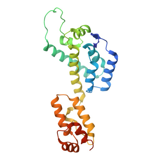
The HIV-1 capsid, composed of capsid (CA) proteins arranged into a conical surface lattice, serves as a critical platform for host-pathogen interactions. Many host cofactors regulate HIV-1 infection by selectively recognizing the higher-order CA lattice rather than individual monomers or capsomers. Among these, Myxovirus resistance protein B (MxB) is a key cellular restriction factor that binds the capsid via its N-terminal fragment containing an arginine-rich motif (RRR), blocking HIV-1 infection at early infection stages. However, the structural basis of this interaction has remained elusive. Here, we present a structural characterization of the CA lattice in complex with the MxB N-terminal fragment using cryo-electron tomography and subtomogram averaging. Combining cryoEM structures with EM-guided all-atom molecular dynamics simulations, we identify residue-specific interactions between MxB and the CA trimer interfaces. Notably, residues 10-20 of the MxB N-terminus adopt an α-helical conformation that stabilizes binding at the CA trimer interface, revealing a previously unrecognized mode of capsid engagement. These findings provide new insight into arginine-rich motif-mediated recognition by MxB and other host factors and establish a structural framework for the development of capsid-targeting antiviral therapeutics.
- Division of Structural Biology, Wellcome Centre for Human Genetics, University of Oxford, Oxford OX3 7BN, UK.
Organizational Affiliation: