9SGE | pdb_00009sge

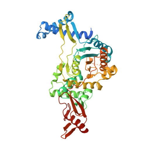
PENICILLIN-BINDING PROTEIN 1B (PBP-1B) IN COMPLEX WITH A MONOBACTAM (Aztreonam)


Experimental Data Snapshot

  • Method: X-RAY DIFFRACTION
  • Resolution: 1.65 Å
  • R-Value Free: 
    0.190 (Depositor), 0.199 (DCC) 
  • R-Value Work: 
    0.173 (Depositor), 0.184 (DCC) 

Starting Model: experimental
View more details

wwPDB Validation 3D Report Full Report

Validation slider image for 9SGE

Ligand Structure Quality Assessment 


This is version 1.1 of the entry. See complete history

Literature

Structure-Activity Relationship and Crystallographic Study of New Monobactams.

Kavas, V.Contreras-Martel, C.Pajk, S.Knez, D.Martins, A.Gould, T.A.Roper, D.I.Zdovc, I.Dessen, A.Hrast Rambaher, M.Gobec, S.

(2026) J Med Chem 69: 3887-3901

  • DOI: https://doi.org/10.1021/acs.jmedchem.5c02427
  • Primary Citation Related Structures: 
    9SG5, 9SG6, 9SG7, 9SG8, 9SG9, 9SGA, 9SGB, 9SGC, 9SGD, 9SGE

  • PubMed Abstract: 

    Monobactams, a subclass of β-lactam antibiotics with a monocyclic scaffold, are uniquely resistant to hydrolysis by metallo-β-lactamases, providing a distinct therapeutic advantage. Here, we report an in silico -based structure-activity relationship (SAR) investigation of aztreonam-related monobactams. A focused library of monobactam derivatives was synthesized and evaluated for inhibition of penicillin-binding proteins (PBPs) and antibacterial activity. Ten compounds, including aztreonam, were crystallized with truncated PBP1b from Streptococcus pneumoniae , used as a model PBP. Potent PBP1b inhibitors were developed, although high enzymatic potency was not always reflected in strong antibacterial activity. Certain derivatives showed activity against Staphylococcus aureus , which is typically resistant to monobactams. 2D similarity search identified potent inhibitors active against Escherichia coli , Klebsiella pneumoniae , and Acinetobacter baumannii . Crystal structures revealed previously unrecognized binding interactions, including a halogen bond with a conserved threonine residue, underscoring the potential of these interactions to support the development of more potent PBP inhibitors.


  • Organizational Affiliation
    • Department of Pharmaceutical Chemistry, Faculty of Pharmacy, University of Ljubljana, Aškerčeva cesta 7, 1000 Ljubljana, Slovenia.

Macromolecule Content 

  • Total Structure Weight: 55.61 kDa 
  • Atom Count: 3,962 
  • Modeled Residue Count: 448 
  • Deposited Residue Count: 494 
  • Unique protein chains: 1

Macromolecules

Find similar proteins by:|  3D Structure
Entity ID: 1
MoleculeChains  Sequence LengthOrganismDetailsImage
Penicillin-binding protein 1B494Streptococcus pneumoniaeMutation(s): 3 
Gene Names: pbp1b
UniProt
Find proteins for O70038 (Streptococcus pneumoniae)
Explore O70038 
Go to UniProtKB:  O70038
Entity Groups
Sequence Clusters30% Identity50% Identity70% Identity90% Identity95% Identity100% Identity
UniProt GroupO70038
Sequence Annotations
Expand
Reference Sequence

Small Molecules

Ligands 3 Unique
IDChains Name / Formula / InChI Key2D Diagram3D Interactions
AZR
(Subject of Investigation/LOI)

Query on AZR


Download:Ideal Coordinates CCD File
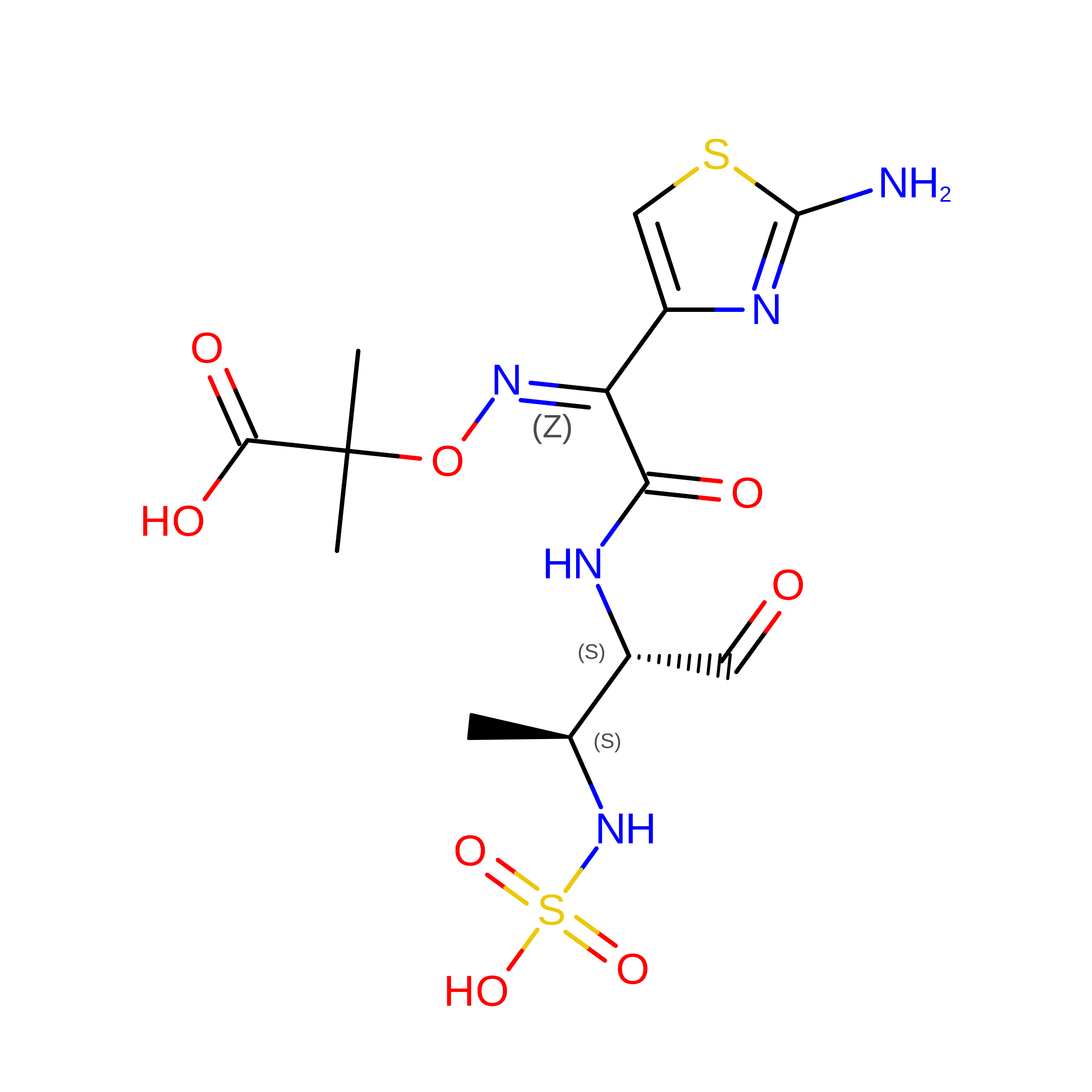
B [auth A]2-({[(1Z)-1-(2-amino-1,3-thiazol-4-yl)-2-oxo-2-{[(2S,3S)-1-oxo-3-(sulfoamino)butan-2-yl]amino}ethylidene]amino}oxy)-2-methylpropanoic acid
C13 H19 N5 O8 S2
OEVQTUTXOVDNJO-GEEOIBTQSA-N
TAU
(Subject of Investigation/LOI)

Query on TAU


Download:Ideal Coordinates CCD File
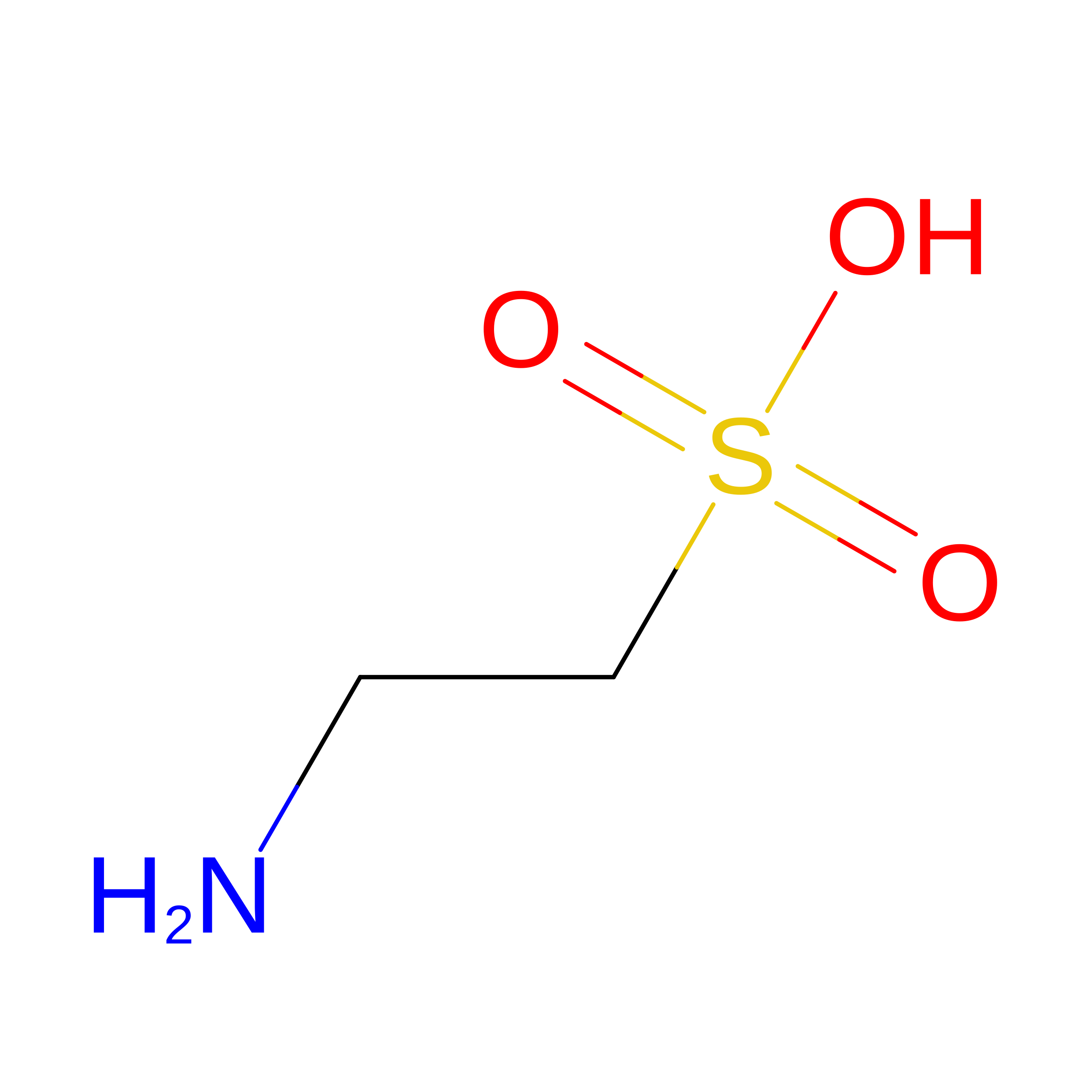
DA [auth A]2-AMINOETHANESULFONIC ACID
C2 H7 N O3 S
XOAAWQZATWQOTB-UHFFFAOYSA-N
CL

Query on CL


Download:Ideal Coordinates CCD File
AA [auth A]
BA [auth A]
C [auth A]
CA [auth A]
D [auth A]
AA [auth A],
BA [auth A],
C [auth A],
CA [auth A],
D [auth A],
E [auth A],
F [auth A],
G [auth A],
H [auth A],
I [auth A],
J [auth A],
K [auth A],
L [auth A],
M [auth A],
N [auth A],
O [auth A],
P [auth A],
Q [auth A],
R [auth A],
S [auth A],
T [auth A],
U [auth A],
V [auth A],
W [auth A],
X [auth A],
Y [auth A],
Z [auth A]
CHLORIDE ION
Cl
VEXZGXHMUGYJMC-UHFFFAOYSA-M

Experimental Data & Validation

Experimental Data

  • Method: X-RAY DIFFRACTION
  • Resolution: 1.65 Å
  • R-Value Free:  0.190 (Depositor), 0.199 (DCC) 
  • R-Value Work:  0.173 (Depositor), 0.184 (DCC) 
Space Group: C 2 2 21
Unit Cell:
Length ( Å )Angle ( ˚ )
a = 96.306α = 90
b = 147.18β = 90
c = 98.117γ = 90
Software Package:
Software NamePurpose
REFMACrefinement
XDSdata reduction
XSCALEdata scaling
PHASERphasing

Structure Validation

View Full Validation Report


Ligand Structure Quality Assessment 


Entry History 

& Funding Information

Deposition Data


Funding OrganizationLocationGrant Number
Slovenian Research AgencySloveniaP1-0208
Slovenian Research AgencySloveniaJ1-50039
Slovenian Research AgencySloveniaJ3-50123
French Infrastructure for Integrated Structural Biology (FRISBI)FranceANR-10-INBS-05-02
Grenoble Alliance for Integrated Structural Cell Biology (GRAL)FranceANR-17-EURE-0003

Revision History  (Full details and data files)

  • Version 1.0: 2026-02-18
    Type: Initial release
  • Version 1.1: 2026-03-11
    Changes: Database references