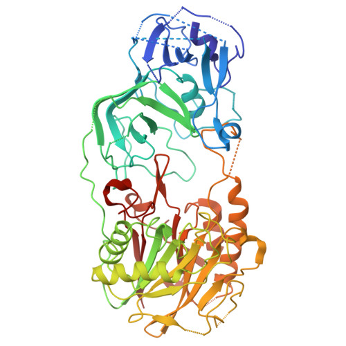
9SF9 | pdb_00009sf9

Dimeric form of the Streptococcus pyogenes nuclease A (SpnA)


Experimental Data Snapshot

  • Method: ELECTRON MICROSCOPY
  • Resolution: 2.80 Å
  • Aggregation State: PARTICLE 
  • Reconstruction Method: SINGLE PARTICLE 

wwPDB Validation 3D Report Full Report

Validation slider image for 9SF9

This is version 1.2 of the entry. See complete history. 

Literature

Molecular mechanism of complement interference by Streptococcus pyogenes nuclease A.

A Bennig, I., Strobaek, J., Mamede, R., Sellerberg, A., Neumann, A., Friaes, A., Ramirez, M., Hall, M., Collin, M., Malmstrom, L., Ekstrom, S., Frick, I.M., Bjorck, L., Happonen, L.J.

(2026) Commun Biol 9

  • DOI: https://doi.org/10.1038/s42003-026-10956-9
  • Primary Citation Related Structures: 
    9SF8, 9SF9

  • PubMed Abstract: 

    Bacteria deploy virulence factors to subvert host immunity; yet the molecular details of these interactions often remain limited. Here, we reveal the structure and host interactome of the Streptococcus pyogenes nuclease A (SpnA). We characterize the structure and dynamics of SpnA using hydrogen-deuterium exchange mass spectrometry and single-particle electron cryo microscopy, yielding the first structural insights to SpnA. This allowed us to identify an additional oligonucleotide-binding domain, whose flexible structure may play an important function in nucleolytic activity. Affinity-pulldown mass spectrometry identified the complement system membrane attack complex (MAC) C5b67 components as key interactors. Cross-linking mass spectrometry combined with integrative modeling identified the binding interfaces between SpnA and C5b67. These interfaces are conserved among genetically diverse S. pyogenes strains. Interaction between SpnA and C5b67 is suggested to prevent the assembly of a functional MAC. Our findings uncover a novel function of SpnA in complement inhibition, and identifies new potential targets to prevent and treat S. pyogenes infections.


  • Organizational Affiliation: 
    • Department of Clinical Sciences, Division of Infection Medicine, Lund University, Lund, Sweden.

Macromolecule Content 

  • Total Structure Weight: 124.18 kDa 
  • Atom Count: 7,560 
  • Modeled Residue Count: 1,075 
  • Deposited Residue Count: 1,126 
  • Unique protein chains: 1

Macromolecules

Find similar proteins by:|  3D Structure
Entity ID: 1
MoleculeChains  Sequence LengthOrganismDetailsImage
Endonuclease/exonuclease/phosphatase domain-containing protein
A, B
563Streptococcus pyogenesMutation(s): 0 
Gene Names: SPy_0747
UniProt
Find proteins for Q9A0J7 (Streptococcus pyogenes serotype M1)
Explore Q9A0J7 
Go to UniProtKB:  Q9A0J7
Entity Groups
Sequence Clusters30% Identity50% Identity70% Identity90% Identity95% Identity100% Identity
UniProt GroupQ9A0J7
Sequence Annotations
Expand
Reference Sequence

Experimental Data & Validation

Experimental Data

  • Method: ELECTRON MICROSCOPY
  • Resolution: 2.80 Å
  • Aggregation State: PARTICLE 
  • Reconstruction Method: SINGLE PARTICLE 
EM Software:
TaskSoftware PackageVersion
MODEL REFINEMENTPHENIX1.21_5207
RECONSTRUCTIONcryoSPARC

Structure Validation

View Full Validation Report


Entry History 

& Funding Information

Deposition Data


Funding OrganizationLocationGrant Number
Swedish Research CouncilSweden2022-03860

Revision History  (Full details and data files)

  • Version 1.0: 2026-09-02
    Type: Initial release
  • Version 1.1: 2026-09-30
    Changes: Data collection, Database references
  • Version 1.2: 2026-10-07
    Changes: Data collection, Database references