9SDJ | pdb_00009sdj

Complex of Ovine Serum Albumin with Hydrolyzed Ampicillin


Experimental Data Snapshot

  • Method: X-RAY DIFFRACTION
  • Resolution: 2.55 Å
  • R-Value Free: 
    0.213 (Depositor), 0.212 (DCC) 
  • R-Value Work: 
    0.164 (Depositor), 0.168 (DCC) 
  • R-Value Observed: 
    0.166 (Depositor) 

Starting Model: experimental
View more details

wwPDB Validation 3D Report Full Report

Validation slider image for 9SDJ

Ligand Structure Quality Assessment 


This is version 1.0 of the entry. See complete history

Literature

Structural Interactions of beta-Lactam Antibiotics with Mammalian Serum Albumins.

Duszynski, K.Sekula, B.Talaj, J.Bujacz, A.

(2026) Int J Mol Sci 27

  • DOI: https://doi.org/10.3390/ijms27020776
  • Primary Citation Related Structures: 
    9Q8U, 9QCH, 9S42, 9S43, 9S73, 9SC5, 9SDJ

  • PubMed Abstract: 

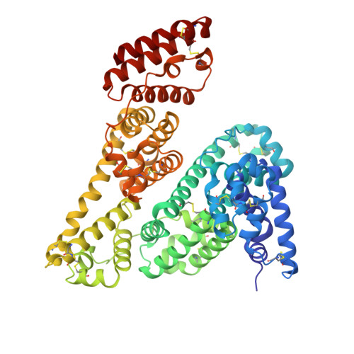
    The Bactericidal action of β-lactam antibiotics is related to covalent modification of transpeptidases, enzymes that take part in the synthesis of bacterial cell wall. The β-lactam moiety mimics the transpeptidase substrate and irreversibly inhibits the enzyme. In penicillin and cephalosporin, the β-lactam ring is coupled with a five-membered thiazolidine ring or a six-membered dihydrothiazine ring, respectively. In the case of penicillins, such conjunction causes higher tension of this bicyclic moiety; therefore, the β-lactam ring can be hydrolyzed in certain conditions, inactivating the antibiotic. Serum albumin is known for its drug binding capabilities, which enable it to transport pharmaceuticals through the circulatory system. Penicillins and cephalosporins are no exception in this aspect, and they are also carried by serum albumin in the bloodstream. In this study, we structurally investigate the ability of three serum albumins-equine (ESA), caprine (CSA), and ovine (OSA)-to bind two penicillins, ampicillin (Amp) and oxacillin (Oxa), and two cephalosporins, cefaclor (Cef) and cephalosporin C (Csc). The crystal structures of these mammalian serum albumin complexes shed new light on the albumin binding properties of β-lactam antibiotics, showing one common binding site for Amp, Oxa, and Cef in Fatty Acid Site 6 (FA6), and a second cefaclor molecule bound in domain I of the equine serum albumin. It was surprising that these antibiotics are not bound in the main drug binding site. However, cephalosporin C is bound in OSA Drug Site 1 (DS1).


  • Organizational Affiliation
    • Institute of Molecular and Industrial Biotechnology, Lodz University of Technology, Stefanowskiego 2/22, 90-573 Lodz, Poland.

Macromolecule Content 

  • Total Structure Weight: 67.77 kDa 
  • Atom Count: 4,804 
  • Modeled Residue Count: 581 
  • Deposited Residue Count: 583 
  • Unique protein chains: 1

Macromolecules

Find similar proteins by:|  3D Structure
Entity ID: 1
MoleculeChains  Sequence LengthOrganismDetailsImage
Serum albumin583Ovis ariesMutation(s): 0 
UniProt
Find proteins for P14639 (Ovis aries)
Explore P14639 
Go to UniProtKB:  P14639
Entity Groups
Sequence Clusters30% Identity50% Identity70% Identity90% Identity95% Identity100% Identity
UniProt GroupP14639
Sequence Annotations
Expand
Reference Sequence

Small Molecules

Ligands 5 Unique
IDChains Name / Formula / InChI Key2D Diagram3D Interactions
ZZ7
(Subject of Investigation/LOI)

Query on ZZ7


Download:Ideal Coordinates CCD File
B [auth A](2R,4S)-2-[(R)-{[(2R)-2-amino-2-phenylacetyl]amino}(carboxy)methyl]-5,5-dimethyl-1,3-thiazolidine-4-carboxylic acid
C16 H21 N3 O5 S
KDAWOPKDXRJNHV-MPPDQPJWSA-N
TLA

Query on TLA


Download:Ideal Coordinates CCD File
F [auth A]L(+)-TARTARIC ACID
C4 H6 O6
FEWJPZIEWOKRBE-JCYAYHJZSA-N
MLI

Query on MLI


Download:Ideal Coordinates CCD File
J [auth A],
K [auth A],
L [auth A],
M [auth A],
N [auth A]
MALONATE ION
C3 H2 O4
OFOBLEOULBTSOW-UHFFFAOYSA-L
ACT

Query on ACT


Download:Ideal Coordinates CCD File
G [auth A],
H [auth A],
I [auth A]
ACETATE ION
C2 H3 O2
QTBSBXVTEAMEQO-UHFFFAOYSA-M
FMT

Query on FMT


Download:Ideal Coordinates CCD File
C [auth A],
D [auth A],
E [auth A]
FORMIC ACID
C H2 O2
BDAGIHXWWSANSR-UHFFFAOYSA-N

Experimental Data & Validation

Experimental Data

  • Method: X-RAY DIFFRACTION
  • Resolution: 2.55 Å
  • R-Value Free:  0.213 (Depositor), 0.212 (DCC) 
  • R-Value Work:  0.164 (Depositor), 0.168 (DCC) 
  • R-Value Observed: 0.166 (Depositor) 
Space Group: P 32 2 1
Unit Cell:
Length ( Å )Angle ( ˚ )
a = 121.189α = 90
b = 121.189β = 90
c = 121.965γ = 120
Software Package:
Software NamePurpose
REFMACrefinement
PDB_EXTRACTdata extraction
XDSdata reduction
XSCALEdata scaling
MOLREPphasing

Structure Validation

View Full Validation Report


Ligand Structure Quality Assessment 


Entry History 

& Funding Information

Deposition Data


Funding OrganizationLocationGrant Number
Polish National Science CentrePoland2013/11/B/T5/02271

Revision History  (Full details and data files)

  • Version 1.0: 2026-02-04
    Type: Initial release