9S6T | pdb_00009s6t

Ternary cryo-EM structure of chicken ALG12 with Dol25-PP-GlcNAc2Man7, Dol25-P-Man, and Fab


Experimental Data Snapshot

  • Method: ELECTRON MICROSCOPY
  • Resolution: 2.42 Å
  • Aggregation State: PARTICLE 
  • Reconstruction Method: SINGLE PARTICLE 

wwPDB Validation 3D Report Full Report

Validation slider image for 9S6T

This is version 1.1 of the entry. See complete history

Literature

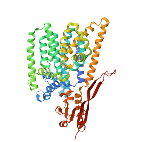
Structures of ALG3/9/12 reveal the assembly logic of the N-glycan oligomannose core.

Alexander, J.A.N.Chen, S.Y.Mukherjee, S.de Capitani, M.Irobalieva, R.N.Rossi, L.Agrawal, P.Kowal, J.Meirelles, M.A.Aebi, M.Reymond, J.L.Kossiakoff, A.A.Riniker, S.Locher, K.P.

(2026) Nat Chem Biol 

  • DOI: https://doi.org/10.1038/s41589-026-02164-7
  • Primary Citation Related Structures: 
    9S6R, 9S6S, 9S6T, 9S6U

  • PubMed Abstract: 

    Asparagine-linked glycans are essential for the maturation and function of most eukaryotic secretory proteins. The biosynthesis and transfer of dolichylpyrophosphate-anchored GlcNAc 2 Man 9 Glc 3 glycan is a highly conserved process occurring in the endoplasmic reticulum (ER) membrane and involving over a dozen membrane proteins whose dysfunction is linked to congenital disorders of glycosylation (CDGs). Three membrane-integral mannosyltransferases, ALG3, ALG9 and ALG12, mediate four consecutive mannosylation reactions that convert GlcNAc 2 Man 5 to GlcNAc 2 Man 9 . Here, using chemoenzymatically synthesized lipid-linked glycan donor and acceptor analogs, we recapitulated this biosynthetic pathway in vitro. High-resolution cryo-electron microscopy structures of pseudo-Michaelis complexes of each step revealed how the branched glycan is accurately synthesized and unwanted side products are averted. Molecular dynamics simulations and mutagenesis studies uncovered a subtle but precise mechanism selecting the dolichylphosphomannose donor substrate over dolichylphosphoglucose, which is also present in the ER membrane. Our results also provide mechanistic explanations for enzyme dysfunction in CDGs and offer opportunities for N-glycan engineering.


  • Organizational Affiliation
    • Institute of Molecular Biology and Biophysics, Eidgenössische Technische Hochschule (ETH) Zürich, Zurich, Switzerland.

Macromolecule Content 

  • Total Structure Weight: 108.76 kDa 
  • Atom Count: 6,007 
  • Modeled Residue Count: 708 
  • Deposited Residue Count: 945 
  • Unique protein chains: 3

Macromolecules

Find similar proteins by:|  3D Structure
Entity ID: 1
MoleculeChains  Sequence LengthOrganismDetailsImage
Mannosyltransferase496Gallus gallusMutation(s): 1 
Gene Names: ALG12
EC: 2.4.1
UniProt
Find proteins for F1P077 (Gallus gallus)
Explore F1P077 
Go to UniProtKB:  F1P077
Entity Groups
Sequence Clusters30% Identity50% Identity70% Identity90% Identity95% Identity100% Identity
UniProt GroupF1P077
Sequence Annotations
Expand
Reference Sequence
Find similar proteins by:|  3D Structure
Entity ID: 2
MoleculeChains  Sequence LengthOrganismDetailsImage
Gg12-11 Fab heavy chainB [auth H]234synthetic constructMutation(s): 0 
Find similar proteins by:|  3D Structure
Entity ID: 3
MoleculeChains  Sequence LengthOrganismDetailsImage
Gg12-11 Fab light chainC [auth L]215synthetic constructMutation(s): 0 

Small Molecules

Ligands 4 Unique
IDChains Name / Formula / InChI Key2D Diagram3D Interactions
A1JMC
(Subject of Investigation/LOI)

Query on A1JMC


Download:Ideal Coordinates CCD File
E [auth A][(2~{R},3~{R},4~{R},5~{S},6~{R})-3-acetamido-5-[(2~{S},3~{R},4~{R},5~{S},6~{R})-3-acetamido-6-(hydroxymethyl)-5-[(2~{S},3~{S},4~{S},5~{R},6~{R})-4-[(2~{R},3~{S},4~{S},5~{S},6~{R})-6-(hydroxymethyl)-3-[(2~{R},3~{S},4~{S},5~{S},6~{R})-6-(hydroxymethyl)-3-[(2~{R},3~{S},4~{S},5~{S},6~{R})-6-(hydroxymethyl)-3,4,5-tris(oxidanyl)oxan-2-yl]oxy-4,5-bis(oxidanyl)oxan-2-yl]oxy-4,5-bis(oxidanyl)oxan-2-yl]oxy-6-[[(2~{S},3~{S},4~{S},5~{R},6~{R})-6-(hydroxymethyl)-4-[(2~{R},3~{S},4~{S},5~{S},6~{R})-6-(hydroxymethyl)-3-[(2~{R},3~{S},4~{S},5~{S},6~{R})-6-(hydroxymethyl)-3,4,5-tris(oxidanyl)oxan-2-yl]oxy-4,5-bis(oxidanyl)oxan-2-yl]oxy-3,5-bis(oxidanyl)oxan-2-yl]oxymethyl]-3,5-bis(oxidanyl)oxan-2-yl]oxy-4-oxidanyl-oxan-2-yl]oxy-6-(hydroxymethyl)-4-oxidanyl-oxan-2-yl] [oxidanyl-[(3~{S},6~{Z},10~{E},14~{E})-3,7,11,15,19-pentamethylicosa-6,10,14,18-tetraenoxy]phosphoryl] hydrogen phosphate
C83 H142 N2 O52 P2
PWYYAJLOGRNTHW-AGFGAJIBSA-N
AV0

Query on AV0


Download:Ideal Coordinates CCD File
G [auth H]Lauryl Maltose Neopentyl Glycol
C47 H88 O22
MADJBYLAYPCCOO-VWHTXWAPSA-N
Y01

Query on Y01


Download:Ideal Coordinates CCD File
F [auth H]CHOLESTEROL HEMISUCCINATE
C31 H50 O4
WLNARFZDISHUGS-MIXBDBMTSA-N
A1JMT
(Subject of Investigation/LOI)

Query on A1JMT


Download:Ideal Coordinates CCD File
D [auth A][(3S,6Z,10E,14E)-3,7,11,15,19-pentamethylicosa-6,10,14,18-tetraenyl] dihydrogen phosphate
C25 H45 O4 P
GYBNOAFGEKAZTA-YGRYZZMUSA-N

Experimental Data & Validation

Experimental Data

  • Method: ELECTRON MICROSCOPY
  • Resolution: 2.42 Å
  • Aggregation State: PARTICLE 
  • Reconstruction Method: SINGLE PARTICLE 
EM Software:
TaskSoftware PackageVersion
MODEL REFINEMENTPHENIX1.21.2_5419
RECONSTRUCTIONcryoSPARC

Structure Validation

View Full Validation Report


Entry History 

& Funding Information

Deposition Data


Funding OrganizationLocationGrant Number
Swiss National Science FoundationSwitzerland310030_196862
Swiss National Science FoundationSwitzerland315230_220007

Revision History  (Full details and data files)

  • Version 1.0: 2026-03-18
    Type: Initial release
  • Version 1.1: 2026-03-25
    Changes: Data collection, Database references